This website uses cookies to ensure you get the best experience on our website.
- Table of Contents
18 Citations 18 Q&As
14 Citations 17 Q&As
8 Citations
14 Citations
Facts about NACHT, LRR and PYD domains-containing protein 3.
Recruitment of proCASP1 to the inflammasome promotes its activation and CASP1- catalyzed IL1B and IL18 maturation and secretion in the extracellular milieu (PubMed:28847925). Activation of NLRP3 inflammasome is also required for HMGB1 secretion (PubMed:22801494).
Human | |
---|---|
Gene Name: | NLRP3 |
Uniprot: | Q96P20 |
Entrez: | 114548 |
Belongs to: |
---|
NLRP family |
AGTAVPRL; AII; AII/AVP; Angiotensin/vasopressin receptor AII/AVP-like; mouse NLRP3; NLRP3; AVP; C1orf7; Caterpiller protein 1.1; CIAS1; CLR1.1; Cold autoinflammatory syndrome 1 protein; Cold autoinflammatory syndrome 1; Cryopyrin; Familial cold autoinflammatory syndrome; FCAS; FCU; Leucine-rich repeat-, and PYD-containing protein 3; Muckle-Wells syndrome; MWS; NACHT; NACHT, LRR and PYD containing protein 3; NACHT, LRR and PYD domains-containing protein 3; NALP3; NLR family, pyrin domain containing 3; NLRP3 mouse; NLRP3 polyclonal; NLRP3 rat; NLRP3; Nucleotide-Binding Oligomerization Domain, Le
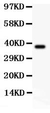
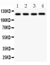
Mass (kDA):
118.173 kDA
Human | |
---|---|
Location: | 1q44 |
Sequence: | 1; NC_000001.11 (247416163..247448823) |
Predominantly expressed in macrophages. Also expressed in dendritic cells, B- and T-cells (at protein level) (PubMed:11786556) (PubMed:17164409). Expressed in LPS-treated granulocytes, but not in resting cells (at protein level) (PubMed:17164409). Expression in monocytes is very weak (at protein level) (PubMed:17164409). Expressed in stratified non- keratinizing squamous epithelium, including oral, esophageal and ectocervical mucosa and in the Hassall's corpuscles in the thymus. Also, detected in the stratified epithelium covering the bladder and ureter (transitional mucosa) (at protein level) (PubMed:17164409). Expressed in chondrocytes (PubMed:12032915). Expressed at low levels in resting osteoblasts (PubMed:17907925).
Cytoplasm, cytosol. Inflammasome. Endoplasmic reticulum. Secreted. Nucleus. In macrophages, under resting conditions, mainly located in the cytosol, on the endoplasmic reticulum. After stimulation with inducers of the NLRP3 inflammasome, mitochondria redistribute in the vicinity of the endoplasmic reticulum in the perinuclear region, which results in colocalization of NLRP3 on the endoplasmic reticulum and PYCARD on mitochondria, allowing the activation of inflammasome assembly. After the induction of pyroptosis, inflammasome specks are released into the extracellular space where they can furt
The NLRP3 marker can be used to study a variety of topics from the cell model that activates NLRP3 to the use of NLRP3 inhibitors to reduce inflammation. These are some of the most important applications of this marker in research. This article will assist you in gaining a more knowledge about the NLRP3 inflammasome, and how to use this biomarker best.
ECs express NLRP3 mRNA and protein which aids in the NLRP3 inflammasome to activate IL-b. Knockdown of NFAT5 reduced both NLRP3 protein and mRNA expression. This suppresses the secretion of mature IL-b by NLRP3 inflammasome-dependent mechanisms. NFAT5 is a key regulator of NLRP3 transcription.
Cholesterol crystals are needed to activate the NLRP3 inflammasome. Cholesterol ester-enriched particle accumulation occurs in vascular endothelial cell lines in mice that have impaired lipoprotein clearance. To investigate the NLRP3-inflammasome's role in the vascular epithelium in the laboratory, the researchers used endothelial cells in culture. To determine the NLRP3 DNA binding sites for the gene they employed bioinformatics analysis and chromatin immunoprecipitation assay.
Activated NLRP3 inflammasomes are expressed in follicular fluids and outside the intrafollicular sac. Inflammasome-mediated gene expression increased and IL-1b production was significantly upregulated after 52-h treatment with PMSG. The researchers speculated that these changes are connected with the ovulation process. Furthermore, NLRP3 inflammasome activity could be a factor in the regulation of ovulation among mammals.
NLRP3 is a cell-specific protein that is found in the perinuclear area, the endoplasmic mitochondria, reticulum, and the nucleus of monocytes that are resting and macrophages. NLRP3 is also found in cytoplasmic accumulations that contain DNA from bacterial sources. It is involved in caspase-1 activationand is activated by a variety of cell damage signals.
In a recent research, NLRP3 inflammasomes were found in ovarian follicles prior to the intrafollicular fluid. These molecules were activated by oocyte maturation during the ovulation. It was found that the quality of oocytes as well as their maturation were closely related and that NLRP3 inflammasome activity was a factor that influenced ovarian follicle development.
In the endothelia of mice consumption of high-salt foods triggers NLRP3 activity. Ingestion of high-salt levels stimulates the expression of IL-1b which then triggers downstream target genes. In mice who ate high salt intake, IL-1b levels were significantly higher than those with a lower salt intake. The effects of high-salt consumption on EC function and the inflammatory response are not fully comprehended.
Atherosclerosis is associated with NLRP3 activation of the inflammasome. OxLDL acts in a similar way to LPS but it also stimulates the release of IL-1b without activating the LPS receptors. In addition the extracellular oxLDL can stimulate the NLRP3 inflammasome through the increase of P2X7R in macrophages.
The development of cancer has been connected to the activation of the inflammasome by tumors. Inflammasome activation influences both adaptive and natural immune responses. It also regulates differentiation, and the microbiota. Many malignancies are linked to the NLRP3 gene which includes colon cancer and Melanoma. Polymorphisms in the NLRP3 gene are linked to an array of inflammation-related diseases, such as cancer, and may even be responsible for the growth of tumors.
Hepatic cancer is the third most prevalent type of cancer that causes the third-highest number of deaths in the world. Its invasiveness is due to activation of the NLRP3 inflammasome. The gene also contributes to liver disease and failure and it has been linked with hepatic stroma. This study also uncovers an inherited polymorphism within the NLRP3 gene that may be linked to the hepatocellular cancer.
Inflammasomes are complexes made up of proteins within the cytosol that play key roles in the initiation of inflammatory responses. The NLRP3 signaling pathway triggers the inflammasome through lysosomal breakdown and reactive oxygen species production. These disruptions could signal the onset or progression of invasive pathogens. Pneumolysin, the most important virulence factor, triggers the disruption of membranes, and causes caspase-1-dependent degradation of the IL-1b gene and IL-18.
The polymorphisms that make up the NLRP3 inflammasome are responsible for activation of the inflammasome in response to different infectious agents. These proteins assist in the contextualization and activation of inflammatory agents by activating the adaptive immune system. However, the mechanisms through which they function are not clear, and further studies are needed to understand their effects in the vivo. There are many promising applications for the inflammasome, including next-generation vaccines.
The NLRP3 inflammasome complex plays an important role in the intestinal epithelium, as evidenced by studies. But, despite the fact that it is associated with human illnesses, NLRP3 inflammasome deficiency in mice can cause increased mortality and loss of body weight. Infection of mice lacking NLRP3 with dextran sodium sulfurate also leads to a greater permeability of the colon, and pathogens lodge in the lymph nodes and liver.
NLRP3 gene polymorphisms have been linked to AIDs, psoriatica and AIDs-related illnesses. Researchers have not found an unambiguous link between NLRP3 gene polymorphisms, AIDs risk, and NLRP3 gene polymorphisms. These associations have been attributed to several factors, including small sample sizes, inadequate statistical power and heterogeneity in disease. Nevertheless, more studies are required to establish the connection between NLRP3 polymorphisms as well as risk for AIDs.
One study revealed that the NLRP3 G-to-C polymorphism significantly increased the risk of suffering from septic shock in patients. Interestingly, the risk was significantly higher for patients who had the GG/GC genotype. The genotype GG/GC was associated with lower survival rates for 28 days and suggests that the NLRP3 G-to-C polymorphism could be possibly a risk factor. Studies of functional analysis have revealed that the 29940G-toC polymorphism is a gain-of function mutation, preventing NLRP3 expression and altering the binding of miR-146a-5p to NLRP3.
The polymorphism 29940 in the NLRP3 gene is associated with susceptibility to sepsis and a poor prognosis. The mutation blocks NLRP3 transcription and the production of inflammatory cytokine downstream. This could lead to new understandings of the pathophysiology behind sepsis and new treatment options for sepsis patients. Although it is restricted to the Han Chinese population it provides new insights into sepsis pathophysiology.
This study is among the first to study the connection between NLRP3 gene polymorphisms and LAA IS. It was found that the T genotype and the T allele were more common in patients suffering from LAA than in controls. This suggests that NLRP3 and CARD8 gene polymorphisms may be linked to susceptibility to LAA. However, there isn't a any significant connection between these genes and LAA.
In addition to the risk of IL-1b CARD8 and NLRP3 are linked to an increased chance of developing autoimmune diseases. CARD8 and NLRP3 are both involved in auto-inflammatory conditions. There is evidence that both genes are linked to a decrease in MAF in Europeans and Polynesians. It has been established that NLRP3 polymorphisms increase the risk for RA and other autoimmune diseases.
The effect of DEX pretreatment on CFs was examined using the CCK-8 assay. Cells pretreated with DEX had a significant reduction in viability. On the other hand, cells treated with H/R had higher levels of viability. MCC950, which is an inhibitor of NLRP3 inflammation, also improved the viability of CF cells when cocultured.
Moreover, mice with the NLRP3 inflammasome-deficient condition were resistant to DSS-induced colitis and were protected from DSS-mediated colitis. Moreover, mice with NLRP3 deficiency were resistant to DSS-induced colitis and were shielded from DSS-mediated colitis. Additionally, the mechanism behind this treatment is to alter NLRP3 expression and reducing the expression of IL-1b in the gut. These signals can also be modulated in patients to offer therapeutic benefits.
NLRP3 is an innate immune system sensor. Two signals trigger the activation of inflammasome. The first signal is generated by bacterial components as well as endogenous cytokines. There are many triggers that trigger the second signal. In the case of activation, it triggers caspase-1 activation and multimerization of the inflammasome. This leads to alarmin being released and pyroptosis.
Dexmedetomidine inhibits NLRP3 expression in cardiac fibroblasts. This is an important source of inflammation in the heart. In this study, dexmedetomidine slowed the activation of the NLRP3 inflammasome and cardiomyocytes showed improved viability and beat rate. The inhibitor also decreased the expression of BAX and Bcl2.
These studies suggest that the VSIG4 gene increases A20 expression and triggers the MS4A6D JAK2-STAT3 signaling pathway. This pathway could be a biomarker or therapeutic target when it is linked to autoinflammatory conditions. Incredibly, Vsig4-/ mice exhibited decreased levels of A20 but this effect did not translate into autoinflammatory disorders that were spontaneously occurring. This may be because the low levels of A20 may control NF-kB activity in macrophages.
NF-kappaB and NLRP3 signaling are crucial in ischemic cardiomyocytes. These cells play a significant role in the functioning of the heart and development. They also play a significant role in MIRI inflammation. The mice's inflammatory response is reduced through the inhibition of inflammation of NLRP3. These inhibitors can be used to reduce MIRI in patients suffering from cardiac fibrosis.
PMID: 11687797 by Hoffman H.M., et al. Mutation of a new gene encoding a putative pyrin-like protein causes familial cold autoinflammatory syndrome and Muckle-Wells syndrome.
PMID: 12355493 by Aganna E., et al. Association of mutations in the NALP3/CIAS1/PYPAF1 gene with a broad phenotype including recurrent fever, cold sensitivity, sensorineural deafness, and AA amyloidosis.
*Showing only the more recent 20. More publications can be found for each product on its corresponding product page