This website uses cookies to ensure you get the best experience on our website.
- Table of Contents
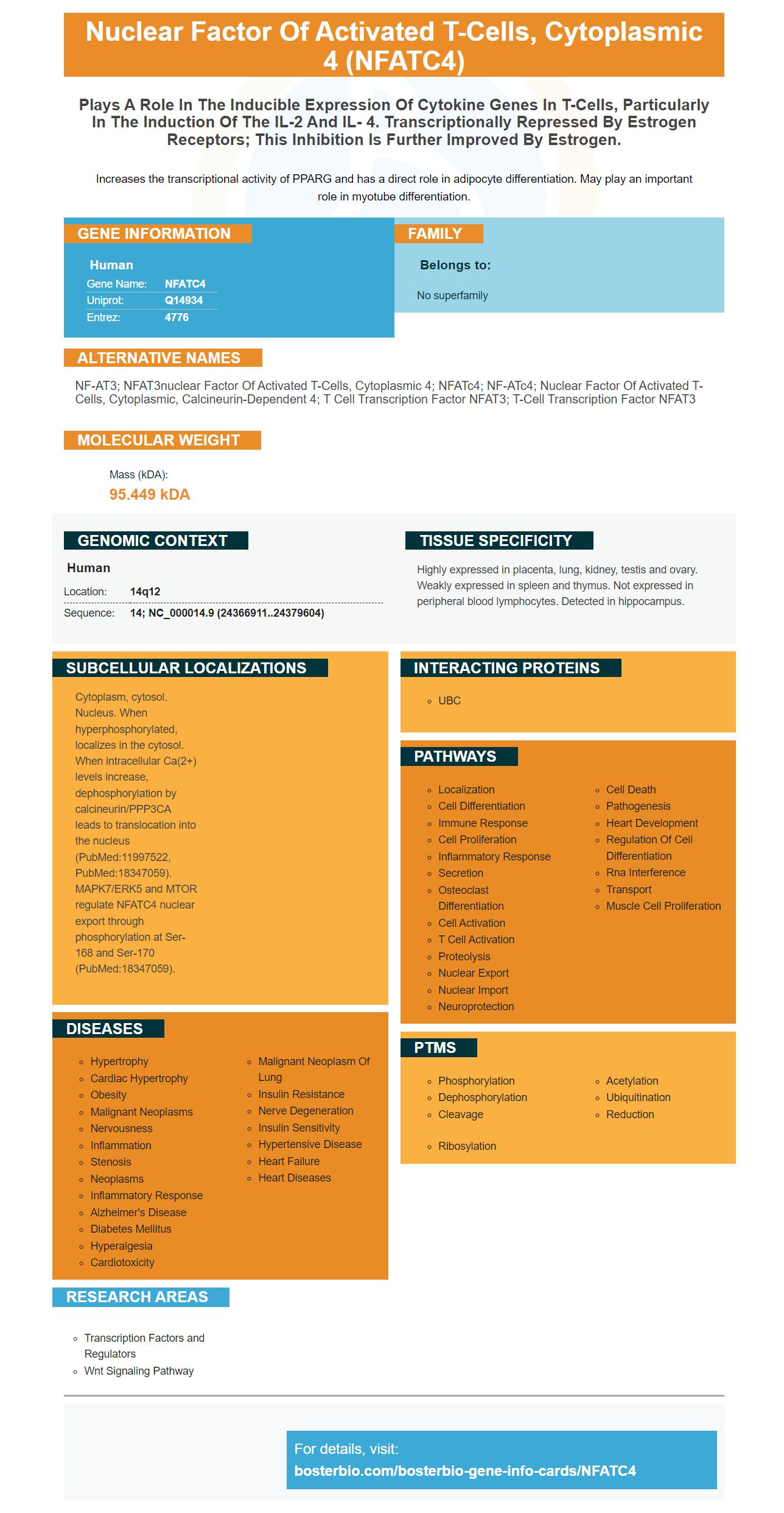


Facts about Nuclear factor of activated T-cells, cytoplasmic 4.

Increases the transcriptional activity of PPARG and has a direct role in adipocyte differentiation. May play an important role in myotube differentiation.
| Human | |
|---|---|
| Gene Name: | NFATC4 |
| Uniprot: | Q14934 |
| Entrez: | 4776 |

| Belongs to: |
|---|
| No superfamily |

NF-AT3; NFAT3nuclear factor of activated T-cells, cytoplasmic 4; NFATc4; NF-ATc4; nuclear factor of activated T-cells, cytoplasmic, calcineurin-dependent 4; T cell transcription factor NFAT3; T-cell transcription factor NFAT3
Mass (kDA):
95.449 kDA

| Human | |
|---|---|
| Location: | 14q12 |
| Sequence: | 14; NC_000014.9 (24366911..24379604) |
Highly expressed in placenta, lung, kidney, testis and ovary. Weakly expressed in spleen and thymus. Not expressed in peripheral blood lymphocytes. Detected in hippocampus.
Cytoplasm, cytosol. Nucleus. When hyperphosphorylated, localizes in the cytosol. When intracellular Ca(2+) levels increase, dephosphorylation by calcineurin/PPP3CA leads to translocation into the nucleus (PubMed:11997522, PubMed:18347059). MAPK7/ERK5 and MTOR regulate NFATC4 nuclear export through phosphorylation at Ser-168 and Ser-170 (PubMed:18347059).


It is vital to employ the right equipment when you conduct experiments in your lab. A Boster Microplate Reader is capable of reading absorbances at 450nm. A Boster Pipette can be used to dispense 0.5 milliliters up to 1 ml of solution in aqueous form. Multichannel pipettes are the best choice for large amounts of samples. Sharing your results can result in product credits.
This boster bio antibody is labeled as A00227-1. It is able to react with Human, Mouse, and Rat. It comes with 5 mg BSA to prepare your sample. The blocking peptide can be purchased for up to one month if looking to do more detailed research. The Boster Bio Anti-GAPDH Antibody Picoband utilizes the NFATC4 marker.
Boster Bio DAB Chromogenic Substrates kit is an instrument that can be used to stain tissue sections and cells. It is used in immunohistochemistry, immunocytochemistry, and cell culture. The buffer is stable at -20°C for one year. Boster Bio DAB Chromogenic Staining Substrate Kit Blue (Blue) is a safe and easy-to-use staining product.
The extracts were separated using 12% SDS-PAGE. The extracts were then electrotransferred onto membranes of nitrocellulose to find out if there was rEgGrx1 within the samples. Membranes were blocked using 5 % (w/v), skim milk. The membranes were incubated using sheep serum, rabbit anti–rEg–Grx1 IgG antibody (1.200 dilution) or goat anti–rabbit IgG. The membranes were then incubated with either a chromogenic substrate, or the Boster Bio DAB ChromogenicSubstrate Kit (Blue).
PMID: 7749981 by Hoey T., et al. Isolation of two new members of the NF-AT gene family and functional characterization of the NF-AT proteins.
PMID: 18675896 by Vihma H., et al. Alternative splicing and expression of human and mouse NFAT genes.


