This website uses cookies to ensure you get the best experience on our website.
- Table of Contents
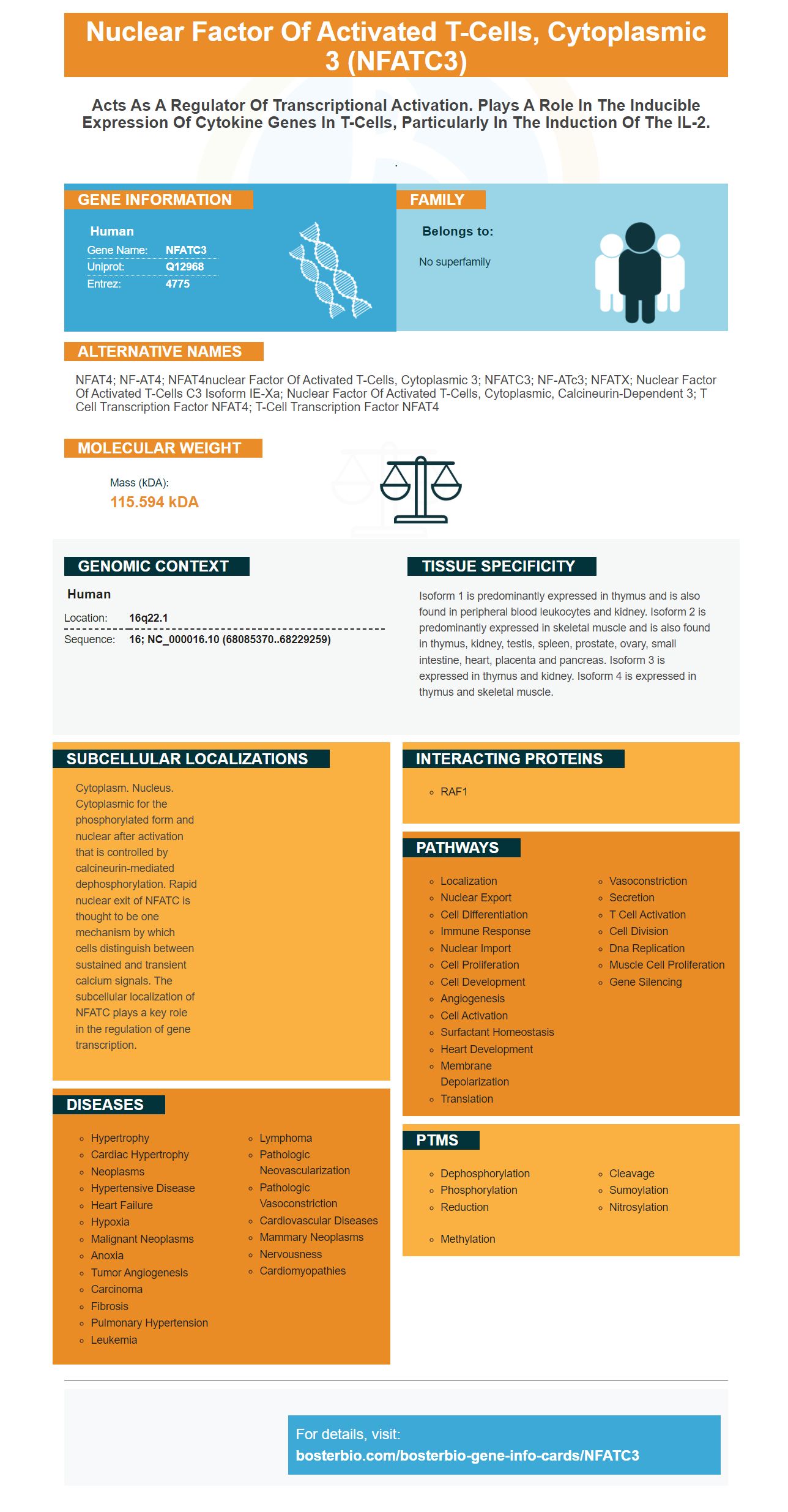
Facts about Nuclear factor of activated T-cells, cytoplasmic 3.
.
Human | |
---|---|
Gene Name: | NFATC3 |
Uniprot: | Q12968 |
Entrez: | 4775 |
Belongs to: |
---|
No superfamily |
NFAT4; NF-AT4; NFAT4nuclear factor of activated T-cells, cytoplasmic 3; NFATC3; NF-ATc3; NFATX; nuclear factor of activated T-cells c3 isoform IE-Xa; nuclear factor of activated T-cells, cytoplasmic, calcineurin-dependent 3; T cell transcription factor NFAT4; T-cell transcription factor NFAT4
Mass (kDA):
115.594 kDA
Human | |
---|---|
Location: | 16q22.1 |
Sequence: | 16; NC_000016.10 (68085370..68229259) |
Isoform 1 is predominantly expressed in thymus and is also found in peripheral blood leukocytes and kidney. Isoform 2 is predominantly expressed in skeletal muscle and is also found in thymus, kidney, testis, spleen, prostate, ovary, small intestine, heart, placenta and pancreas. Isoform 3 is expressed in thymus and kidney. Isoform 4 is expressed in thymus and skeletal muscle.
Cytoplasm. Nucleus. Cytoplasmic for the phosphorylated form and nuclear after activation that is controlled by calcineurin-mediated dephosphorylation. Rapid nuclear exit of NFATC is thought to be one mechanism by which cells distinguish between sustained and transient calcium signals. The subcellular localization of NFATC plays a key role in the regulation of gene transcription.
This Boster Bio Anti-GAPDH Rabbit Monoclonal Antibody Picoband review will focus on its application in Immunofluorescence and In Vitro Diagnostics. Before deciding whether to purchase this product, it's worth considering its features and capabilities. This Boster bio product is validated on multiple platforms with known positive and negative samples to determine its specificity and affinity. In addition, Boster recognizes and rewards scientists who review products and submit them for validation, offering product credits to the first reviewer.
The name of this product is taken from the Chinese character for "mei zhong" - Mei Zhong Chan Pin Jun. In fact, it's better known as "Sui Shi Huan". A recent study by Steven Xia Yu and colleagues found that this antibody can effectively block GAPDH activity in the body. This new antibody is a great way to test your immune system's ability to destroy GAPDH and other proteins that cause aging.
The company's products are characterized by their high quality and excellent validation. They are produced in-house from the highest quality monoclonal antibodies. In addition to that, Boster also offers various conjugates. These include goat anti-rabbit monoclonal antibodies and human polyclonal antibodies. The company's Anti-GAPDH Rabbit Monoclonal Antibody Picoband has been validated on known positive and negative samples.
Boster Bio Anti-GAPDH Rabbilololol is a high-sensitivity, highly specific antibody for the detection of GAPDH. It has been validated on a panel of 250 human, mouse, and rat tissues. Its antibodies have high affinity and are tested against the same quantity of recombinant protein. These products have been used in research for two and a half decades. Boster Bio's antibodies are validated using IHC, WB, ELISA, and flow cytometry.
GAPDH is a surface-located molecule that has multiple applications in biomedical research. Its interactions with plasminogen can help determine the cause of disease. In addition to being surface-located, GAPDH also acts as a plasminogen receptor. It also helps break down the extracellular matrix. And there are many other applications for this antibody.
The expression of the NFATC3 gene is required for glioma migration and proliferation, and knockdown of the gene inhibits the processes. We have used transwell assays to test the effects of knockdown on cell proliferation and migration. The expression of NFATc3 in gliomas resembles that of the disease in vivo. To study the role of this gene, we have used transwell assays with cells incubated in 2% FBS. We then performed flow cytometry on these cells at 1 and 3 days after transfection. The dilution of the fluorescent compound was observed in every division.
We used a knockout mouse model to determine the expression of NFATc3 and NFATc4 in neuronal cells. We used the NFATc3 antibody to label neurons and use a CN-dependent NFATc dephosphorylation inhibitor, CsA, to prevent the proteins from interacting with the luciferase label. The luciferase activity was then measured in these cells in "in vivo" by measuring bioluminescence intensity.
Lithium-treated mice showed increased levels of NFATc4 and NFATc3 nuclear staining. In addition, DAPI-nuclear counterstaining allowed us to confirm the NFATc4 nuclear localization. Finally, CsA prevented lithium-induced apoptosis and motor deficits. We have further used this marker in immunohistochemistry.
The nuclear NFATc3 gene has been implicated in cancer growth. NFATc3 is known to regulate the expression of RCAN1-4, GM-CSF, IL-2, and CXCR-3. In addition, NFATc3 overexpression did not enhance IL-8 induction in these cells. Moreover, the overexpression of NFATc3 did not promote tumour growth in PMA-stimulated cells.
The NFATc3 gene is highly expressed in muscle cells and lymphoid tissues. Besides, it has been described as a negative regulator in muscle cells. This finding is a significant step forward in advancing the science of immunofluorescence. Although there are still some challenges, we have found a new marker that can help us in our research. We are excited to share this new tool with the world!
ATF3 is a member of the ATF/cAMP response element-binding protein family. It can regulate gene activity and translocate to the nucleus. It also has a role in regulating the expression of Wnt6. Our results show that ATF3 is an important player in podocyte injury and repair. We are excited about the new research into the role of this gene.
The Boster Bio NFATC3 ELSA Kit is an assay for the detection of the protein NFATC3. It is a human molecule encoded by the gene NFATC3. The protein is part of the nuclear factors of activated T cells (NFAT) DNA-binding transcription complex, and it regulates the expression of genes in T and immature thymocytes. The kit is validated to detect NFATC3 in 96 samples.
The NFATC3 marker was originally identified as a novel marker for pulmonary disease. It was discovered that this gene can be detected in respiratory epithelial cells at E17.5 and E18.5. In the present study, Nfatc3 was found to interact with the Abca3 gene and is expressed in MLE-15 cells. To validate this marker for lung disease, the mouse Gapdh promoter was used as a control and input DNA served as a positive control. The colocalization of Nfatc3 with TTF-1 was detected using yellow fluorescence.
The expression of this gene is consistently reduced in failing hearts, mouse and rat cardiomyocytes. Nuclear factor of activated T cells (NFAT) pathway suppression was identified by signal pathway screening. NULP1 binds to the topologically associating domain of NFAT3. Moreover, inactivation of the NFAT pathway rescued pathogenesis of cardiac hypertrophy.
PMID: 7749981 by Hoey T., et al. Isolation of two new members of the NF-AT gene family and functional characterization of the NF-AT proteins.
PMID: 7739550 by Masuda E.S., et al. NFATx, a novel member of the nuclear factor of activated T cells family that is expressed predominantly in the thymus.