This website uses cookies to ensure you get the best experience on our website.
- Table of Contents
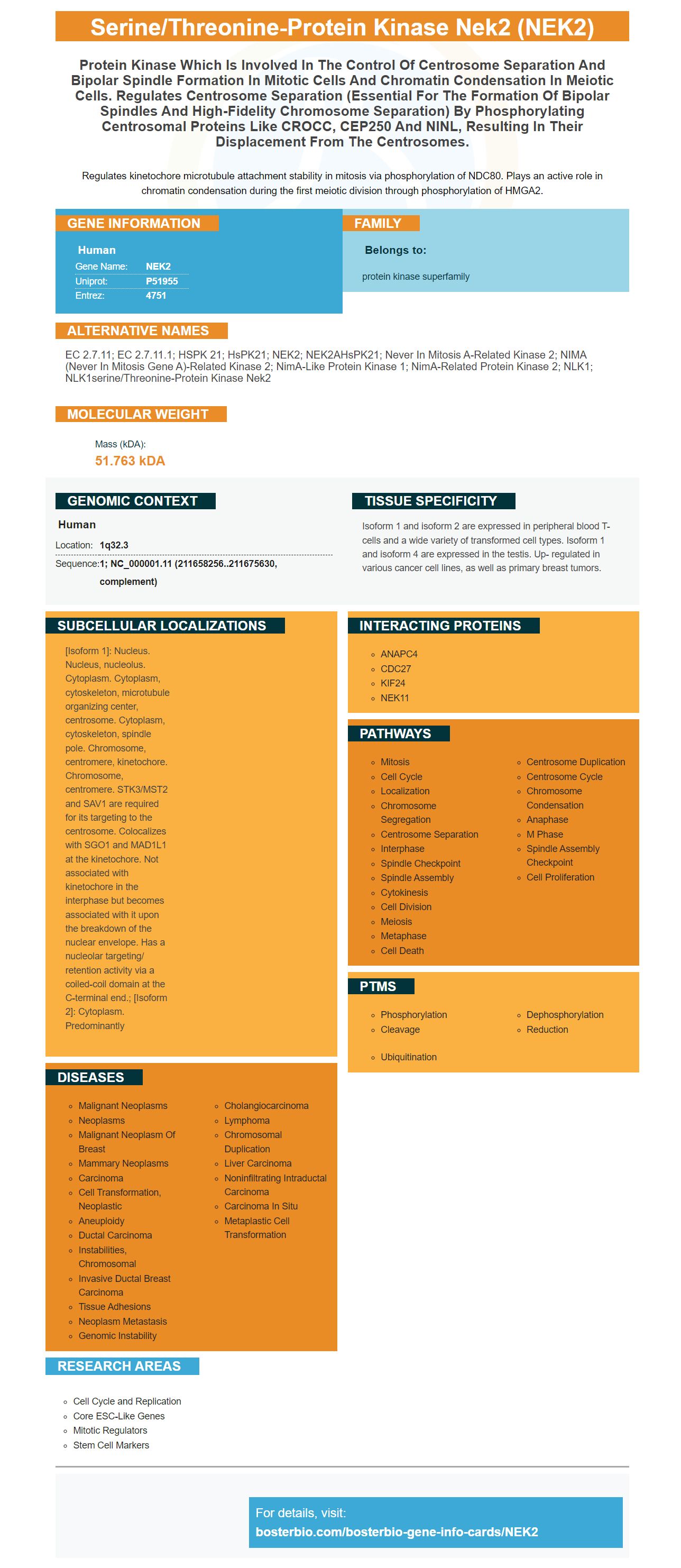
Facts about Serine/threonine-protein kinase Nek2.

Regulates kinetochore microtubule attachment stability in mitosis via phosphorylation of NDC80. Plays an active role in chromatin condensation during the first meiotic division through phosphorylation of HMGA2.
| Human | |
|---|---|
| Gene Name: | NEK2 |
| Uniprot: | P51955 |
| Entrez: | 4751 |

| Belongs to: |
|---|
| protein kinase superfamily |

EC 2.7.11; EC 2.7.11.1; HSPK 21; HsPK21; NEK2; NEK2AHsPK21; Never in mitosis A-related kinase 2; NIMA (never in mitosis gene a)-related kinase 2; NimA-like protein kinase 1; NimA-related protein kinase 2; NLK1; NLK1serine/threonine-protein kinase Nek2
Mass (kDA):
51.763 kDA

| Human | |
|---|---|
| Location: | 1q32.3 |
| Sequence: | 1; NC_000001.11 (211658256..211675630, complement) |
Isoform 1 and isoform 2 are expressed in peripheral blood T-cells and a wide variety of transformed cell types. Isoform 1 and isoform 4 are expressed in the testis. Up- regulated in various cancer cell lines, as well as primary breast tumors.
[Isoform 1]: Nucleus. Nucleus, nucleolus. Cytoplasm. Cytoplasm, cytoskeleton, microtubule organizing center, centrosome. Cytoplasm, cytoskeleton, spindle pole. Chromosome, centromere, kinetochore. Chromosome, centromere. STK3/MST2 and SAV1 are required for its targeting to the centrosome. Colocalizes with SGO1 and MAD1L1 at the kinetochore. Not associated with kinetochore in the interphase but becomes associated with it upon the breakdown of the nuclear envelope. Has a nucleolar targeting/ retention activity via a coiled-coil domain at the C-terminal end.; [Isoform 2]: Cytoplasm. Predominantly


This article will explain the NEK2 Marker and how it is used in Boster Bio. This article will explain how this unique marker works at Boster Bio and how you can use it in your own experiments. NEK2 can be used as a phosphorylated NEK2 and is a powerful tool to researchers.
The NEK2 gene is a transcription factor that facilitates the epithelial-to-mesenchymal transition (EMT). Studies have shown that overexpression of Nek2 is associated with increased expression of EMT markers such as vimentin and SNAIL. These studies don't prove that Nek2 causes breast cancer. However they do show that the gene is crucial in the progression of the disease.
The NEK2 genes is a key regulator for various enzymes. It is necessary for a variety of metabolic processes. The NEK2 gene does not exist in all cells and is especially important in cancer. It is crucial to study the NEK2 genetic code as NEK2 expression varies greatly depending on cell types. We will now discuss how you can use this gene to your advantage.
Overexpression of NEK2 in mice is associated with poor survival rates and disease-free survival. The expression of NEK2 correlates with centrosome proteins implicated in cancer, including CEP55, CENPA, and the CIN gene. These associations suggest that NEK2 could be used as a prognostic marker in lung cancer and as a therapeutic target in adenocarcinoma.
NEK2 acts as a regulator of cell growth. This protein promotes cell proliferation by promoting the transition from the S to the G2/M phases. HCC cells are dependent on NEK2. However, a negative control for Nek2 was performed by using MBP. The early detection of the disease is possible by monitoring NEK2 activity. HCC does not have a cure. However, there are many ways to monitor the disease's progression.
Nek2 expression was identified using a northern blot analysis of several human cell lines. Several human cell lines express Nek2 at higher levels. Consequently, NEK2 makes a great target for cancer intervention. It can be targeted by RNA interference. This means that it blocks the ATP binding area or interferes with protein interactions. Further research is needed in order to fully understand Nek2.
In the past, NEK2 has been linked to the progression of various types of cancer. This protein has also shown to be highly expressed in a variety human cancers. NEK2 is a highly expressed protein in human primary tumors. Its overexpression is associated with tumor progression and malign transformation. This protein has many uses. This gene is an important addition to cancer research.
NEK2 is also involved in the regulation of cell division. C-Nap1 is a centrosomal Nek2-associated Protein that Nek2 phosphorylates. This phosphorylation process results in the loss of centrosome-centriole cohesion. The normal function may also be affected if NEK2 is overexpressed.
We were able use immunocytochemistry and Image Pro Plus 6.0 to quantify the NEK2 gene expression level in cell culture samples. This protein is expressed at levels up to 2.21 mg/mL. It can be used in immunocytochemical experiments to study the molecular basis of chromatin condensation. This marker can also be used in research to diagnose disease.
The maintenance of chromatin in meiotic cells is a function of the NEK2 gene. Hence, it is essential for the avoidance of a second round of DNA duplication. Although the molecular targets are unknown for NEK2, it is known that it interacts with HMGA2, an architect chromatin protein. Consequently, HMGA2 levels in cells are decreased when Nek2 is phosphorylated.
Anti-NEK2 mAb was developed against a prokaryotic gene expression system. This antibody is high in affinity, specificity, as well as potency. This monoclonal antibody is used for detection of liver cancer in high-risk populations and may be a useful tool in the fight against the disease. A diagnostic kit for hepatocellular carcinoma can also be developed using antibodies against NEK2.
The NEK2 Gene was identified using a method called amplification and analytic. A full-length NEK2 gene from a human was cloned (HL7702). The target gene of interest was amplified via PCR, and digested with BamHI/Sa1I. The recombinant NEK2 protein was then transfected into E. coli BL21 clones using a pET30a plasmid. In a subsequent step, the cells were induced to produce a positive clone using isopropyl-b-D-thiogalactopyranoside.
A recent study published by J Comput Biol identifies NEK2 in a new biomarker to detect head and neck squamous carcinoma. Researchers discovered that NEK2 could regulate centromere protein and cause abnormal cell division. Additionally, the overexpression of Nek2 leads to the proliferation of multinucleate cells.
Ewing sarcoma first revealed the Nek2 protein. Later, it was discovered that the gene is overexpressed within various types of cancer, including cholangiocarcinoma as well as non-Hodgkin lymphoma. It has also been found in breast, testicular and colon cancers, which may indicate its role as a regulator of cell proliferation. The Nek2 Protein was found in approximately 60% of surgically resected colon carcinoma specimens. It was also detected in 30% of paracancerous specimens and 10% in normal tissue.
In addition to a positive clonal bacterial result, the NEK2 genes were successfully isolated and sequenced. This allowed for the construction of a recombinant vector. The sequence of NEK2 targets gene is identical to that of the clonal bacterial solutions (Additional File 1: Data S1).
The NEK2C gene is identified in boster bio. The corresponding boster bio protein is also identified by its binding ability to microtubules. It can also be found in centrosomes. The Nek2C marker has the capacity to colocalize with g-tubulin and phosphorylates only in cells containing Nek2.
Nek2 is a cell cycle regulated serine/threonine protein kinase. It has been implicated with centrosome separation, bipolar spinningle formation, andchromatin condensation. There are two major Nek2A/B splice variations: Nek2C and Nek2B. The former has an 8-amino acid sequence internal and occurs at the exact same splice site as Nek2A/Nek2B.
NEK2 proteins were purified using a Prokaryotic Expression System and then subjected for immunocytochemical stained. Image-Pro Plus 6.0 software measured the expression levels. Additionally, the purified NEK2 RNA protein concentration was 2.21mg/mL. The protein's exact concentration was not known, as the NEK2 genes is not expressed in all cells.
NEK2 is a member of the third family of mitotic kinases. It regulates cell division and cell cycle progression. Its activity peak during the S and G2 phases. It is involved in the development of many types of cancer. It can also inhibit cell proliferation and tumorigenesis. Anti-NEK2 mAbs can be a viable treatment for liver carcinoma.
This study used a monoclonal anti-NEK2 mAb that was generated in the same way as NEK2 from human serum. The monoclonal antibody was isolated and serially diluted at 1:1024,000. It was incubated at 1:1024,000 with Nek2 monoclonal mouse anti-human antibody. Then, it was washed. The mAb was used as a control and incubated for 1% BSA. The Western Blot was then repeated.
When the mAbs were analyzed it was discovered that a majority were positive for NEK2 protein. These six kinases were found in the top ten predicted kinases according to a statistical analysis. They were further validated with experiments to confirm if NEK2 could be a valid p53 target. The results showed that NEK2 is a valid marker of p53 kinase activity.
PMID: 7522034 by Schultz S.J., et al. Cell cycle-dependent expression of Nek2, a novel human protein kinase related to the NIMA mitotic regulator of Aspergillus nidulans.
PMID: 11742531 by Hames R.S., et al. Alternative splice variants of the human centrosome kinase Nek2 exhibit distinct patterns of expression in mitosis.


