This website uses cookies to ensure you get the best experience on our website.
- Table of Contents
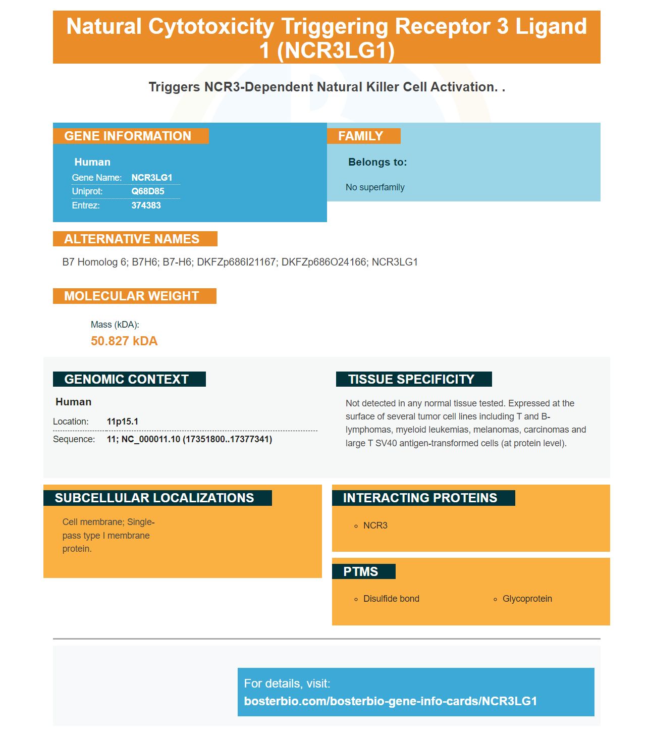
Facts about Natural cytotoxicity triggering receptor 3 ligand 1.
Human | |
---|---|
Gene Name: | NCR3LG1 |
Uniprot: | Q68D85 |
Entrez: | 374383 |
Belongs to: |
---|
No superfamily |
B7 homolog 6; B7H6; B7-H6; DKFZp686I21167; DKFZp686O24166; NCR3LG1
Mass (kDA):
50.827 kDA
Human | |
---|---|
Location: | 11p15.1 |
Sequence: | 11; NC_000011.10 (17351800..17377341) |
Not detected in any normal tissue tested. Expressed at the surface of several tumor cell lines including T and B-lymphomas, myeloid leukemias, melanomas, carcinomas and large T SV40 antigen-transformed cells (at protein level).
Cell membrane; Single-pass type I membrane protein.
This article will talk about NCR3LG1 markers and Boster Bio. We'll also discuss how to use them in your own research. In addition, we'll discuss how primary antibodies are used to identify NK cells. We'll then discuss some other NK marker markers and how these work in biological assays. We'll also discuss the Boster Bio method, which uses a mouse and rabbit to develop antibodies for this marker.
The NCR3LG1 gene marker is a powerful tool for NK cell research. The NCR3 gene regulates a variety of molecules that activate the NK cell response. The target gene CD274 inhibits the function of T cells, while CD283 promotes cytotoxicity. Although the NCR3LG1 is not a candidate for NK cell therapy, it may prove useful in the treatment of cancer patients with cancer.
The NCR3LG1 gene is expressed on many human cells. The gene encodes NCR3LG1 as well as NKCR1. These proteins regulate many cytokine dependent processes. These molecules regulate the translation and transcription of host genes. Their expression is however variable between species. It is therefore important that you determine the target gene of each cell type.
NK cells that express CD56/CD16 can be crucial in viral infection. NK cells that express CD16 are crucial in the ADNKA process. Consequently, improving the gating strategy to include these cells is critical for targeting viral infections. There are many factors that can influence NCR3LG1 expression in human cancer cells. For example, viral CPE can influence the immunological synapse. This could explain why CD56/CD16+ NK cells are important for cancer immunotherapy.
T cell function is inhibited by TNFRSF1B, and MDSC-mediated immunity suppression is supported by TNFR2. NCR3LG1 promotes normal NK cell function. It is remarkable that standard cancer therapies can upregulate the expression of B7/H6. This increases the tumor's sensitivity towards NK cell cytolysis.
A membrane marker is the NK cell antigen CD56brightCD16lo/. It is also a component of cytokines. Different markers are used to identify subsets. The CD56brightCD16+ subset of conventional NK cell population makes up approximately 5-10% circulating peripheral blood. The CD56dim population is more abundant in secondary lymphoid tissues, whereas CD56bright and CD56dim are both expressed in large numbers on NK cells.
By density centrifugation, a pool of PBMCs was obtained from healthy volunteers. PBMCs were incubated at 37°C for 4 h with monensin or CD107 mAb. The cells were then washed in PBS containing 2 M EDTA and stained by a variety of extracellular marker. After PBMCs were fixed in 2% paraformaldehyde solution and permeable using Perm/Wash, they were labeled as CD3+CD56+. APC–IFN–g (BD), detected intracellular IFN–g.
Using the NK Cell Isolation Kit, human PBMCs can be processed and pre-enriched for NK and NKT cells. The NCR3LG1 marker can be used to label non-target cells and magnetically remove them. This results in pure NK cell. The cell population containing NK and NKT cells is enriched in CD56 subsets. This subset is also characterized with a higher percentage of CD56+ cells.
Human natural killer cells are unique lymphocytes with immune-modulating and cytotoxic abilities. They comprise approximately five to ten percent of circulating lymphocytes. They are found in peripheral tissues. They are also known as cytotoxic granules or inflammatory cytokines. These cells are essential in the immune system and play an important role in immunotherapy and cancer biology.
Detecting NK cell presence with primary antibodies is a powerful method. They are widespread throughout tissues and make up a relatively small fraction of the total lymphocyte population, ranging from 2 percent in the mouse spleen to 18% in the human peripheral blood. Their turnover rate is approximately 2 weeks. NK cells are found in a variety of organs including the skin, lungs and liver. They account for twenty to thirty percent all lymphocytes.
A good reagent should recognize granzymeB (an enzyme found in the lysosomal granules and natural killer cell cells) to detect NK-cells. This enzyme, also known as CTLA-1 (cytotoxic T lymphocyte-associated serine esterase), is a crucial factor in the apoptotic pathway of these cells. These enzymes are also important for the immune response and have been implicated in a number of age-related diseases.
Numerous studies have shown human NK cells are sensitive to the CD16 ligand. It is an inhibitory cytokine which triggers cell deaths. Despite having high levels of this receptor expression, the cell is unable or unwilling to cytolyze target cell without the presence of immune cells. In addition, NK cells have the ability to distinguish between'self' and target cells by expressing a low-affinity Fc receptor called CD16.
The use of anti-CD20 mAb ab36388, which binds to the gamma-delta-TCR T cell, did not affect the ADCC of anti-CD20 mAbs, but significantly inhibited the effects of IL-2 on rat NK cells. Abcam also offers knockout edited cell lines and gold-standard validation of its anti-CD20 mAbs.
Purified PBMCs of patients were stained with different antibodies to determine the identity NK cells. The NKp46 marker was used to identify NK cells. Other antibodies were also used in the identification of NK cells such as CD45RA, CD19. These markers allow researchers the ability to compare individual NK-cell counts to their healthy counterparts.
The CD56bright subset is thought to be an earlier stage in NK maturation and features increased proliferative activity. It also contains significant amounts of the key protein, IFNG. CD56dimNK cells, on the other hand, are the result a downregulation CD56. Their expression is associated high levels the cell surface antigen CD16. These cells display cytotoxic activity and contain several markers that bind immunoglobulin G.
These secondary antibodies were used to identify NK cells. PBMCs were fixed-permeable in 50 mls of BD Perm Wash solution containing an anti-GzB–Alexa Fluor 700 antibody cocktail and anti-Ki67-V450 antibody cocktail. The cells were then rinsed twice and resuspended using Staining Buffer. The images were gated with forward scatter (FS), side scatter (SSC), and CD56 versus the CD3 expression.
Secondary antibodies can be used to identify NK cell types. This is a crucial tool in cancer immunosurveillance. These immune cells function as cytolytic effector lymphocytes and directly induce the death of tumor cells. Furthermore, these cells have the ability to produce immune cytokines despite being unspecifically-specific. T cells cannot recognize pathogens that are not present on their cells, but they can detect tumor cells by expressing cytokines when there is a secondary antibody.
PMID: 19528259 by Brandt C.S., et al. The B7 family member B7-H6 is a tumor cell ligand for the activating natural killer cell receptor NKp30 in humans.
PMID: 21422170 by Li Y., et al. Structure of the human activating natural cytotoxicity receptor NKp30 bound to its tumor cell ligand B7-H6.