This website uses cookies to ensure you get the best experience on our website.
- Table of Contents
1 Citations 5 Q&As
Facts about DNA mismatch repair protein Msh2.
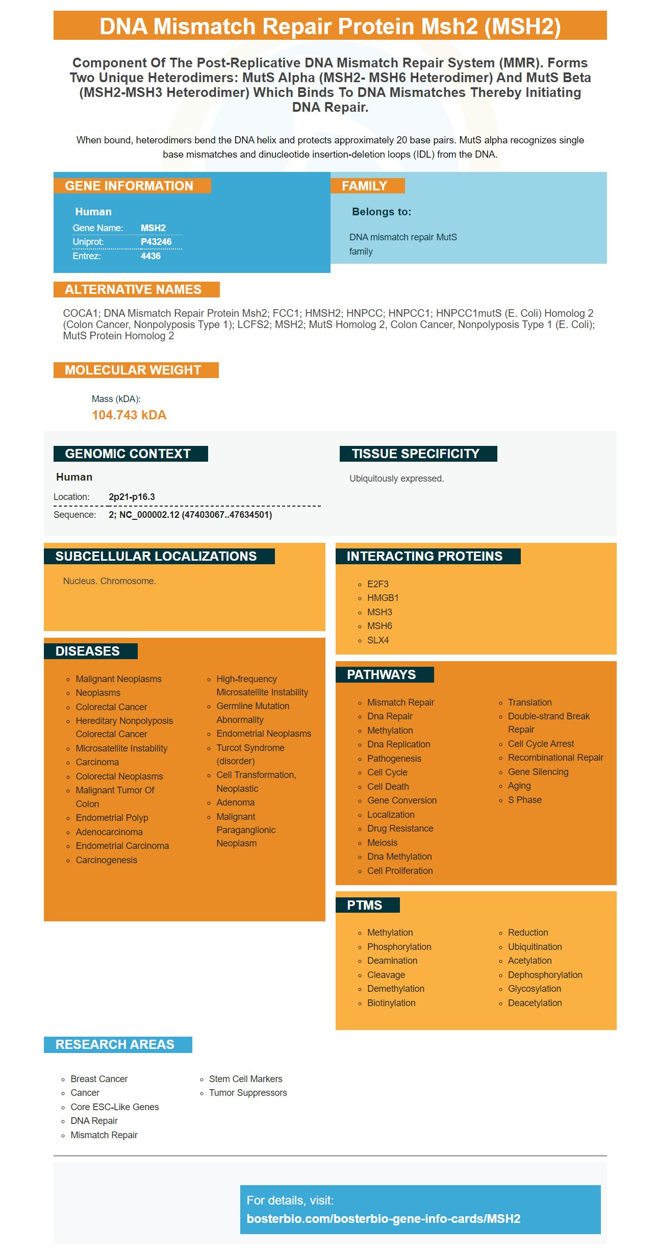
When bound, heterodimers bend the DNA helix and protects approximately 20 base pairs. MutS alpha recognizes single base mismatches and dinucleotide insertion-deletion loops (IDL) from the DNA.
Human | |
---|---|
Gene Name: | MSH2 |
Uniprot: | P43246 |
Entrez: | 4436 |
Belongs to: |
---|
DNA mismatch repair MutS family |
COCA1; DNA mismatch repair protein Msh2; FCC1; hMSH2; HNPCC; HNPCC1; HNPCC1mutS (E. coli) homolog 2 (colon cancer, nonpolyposis type 1); LCFS2; MSH2; mutS homolog 2, colon cancer, nonpolyposis type 1 (E. coli); MutS protein homolog 2
Mass (kDA):
104.743 kDA
Human | |
---|---|
Location: | 2p21-p16.3 |
Sequence: | 2; NC_000002.12 (47403067..47634501) |
Ubiquitously expressed.
Nucleus. Chromosome.
In this course, we will use two unique reagents for MSH2 PCR. One is a template DNA with MSH2 and the other is mutagenic oligonucleotides. Before the course, we constructed pMSH2 template DNA, which we'll discuss in detail in the following section. Both are designed to increase the expression of MSH2 in various cell types.
MutSa mismatch repair subunits are formed by recombination of two genes pGBD and pGSH2. In this study, a two-hybrid reporter strain was transformed with pGBD-MSH2. The diploid cultures were spotted onto Msh2 mutant yeast cells. Assays were carried out by quantifying the nuclear/cytoplasmic fluorescence and determining the presence or absence of Msh2.
The putative pGBD-MSH2 NS cleavage site was identified as codons 525 and 552 in MATa yeast. The cleavage site was identified by sequencing the resulting pGBD-MSH2-MSH2 cDNA. A second set of MATa yeast 2-hybrid reporter strains was used for the analysis.
The cleavage site of the pGBD-MSH2 cDNA was determined by using a strain lacking tryptophan and leucine. The Msh2 variants used were the pMSH6-RFP (Red Fluorescent Protein) cDNA clone. The double NLS mutant protein and Msh2 missense variant were used as negative controls.
The kanamycin marked deletions of MSH3 and MSH6 in W303 RAD5 CAN1 strains were generated by single-step PCR mediated gene disruption. The mutagenic primers used for pGBD-MSH2 cDNA were MSH3R1 and MSH6F2+. PCR experiments with pMR5484 were also performed using the modified kanamycin marked fusion locus.
In addition to the cleavage site, Msh2 protein also exhibited nuclear localization. The presence of Msh3 or Msh6 in cells without Msh6 should prevent Msh2 from nuclear import. Yet, cells lacking both Msh3 and Msh6 still contained Msh2 in the nucleus. This suggests that MutSb is not required for Msh2 nuclear import.
The anti-Integrin alpha 3/ITGA3 antibody from Boster Bio is part of their Picoband(tm) catalog. It has been tested in a variety of applications in flow cytometry and immunohistochemistry. This product reacts with Rat, Mouse, and Human cells. Its trehalose based composition allows it to bind to mouse and human cell lines.
MutSa and MutSb are mismatch repair model organisms. MutSa is a mismatch recognition protein that has two heterodimer partners, MutSb and Msh2. In this heterodimer, the Msh2 and Msh6 molecules cooperate in scanning the genome for insertion/deletion loops. In addition to Msh2 and Msh6, MutSa and Msh6 are required for eukaryotic cell growth and function.
The MutSa to MutSb ratio in a cell is maintained by a regulatory mechanism. It is important to have sufficient coverage in the cell genome and the ratio of monomers in the cytoplasm is correct. Besides maintaining genome coverage, MutSa maintains the stoichiometry of the two subunits in the cell. The MutSa and MutSb homodimers must be responsive to environmental conditions.
PMID: 8252616 by Fishel R., et al. The human mutator gene homolog MSH2 and its association with hereditary nonpolyposis colon cancer.
PMID: 8261515 by Leach F.S., et al. Mutations of a mutS homolog in hereditary nonpolyposis colorectal cancer.
*More publications can be found for each product on its corresponding product page