This website uses cookies to ensure you get the best experience on our website.
- Table of Contents
3 Citations 3 Q&As
3 Citations
Facts about Matrix metalloproteinase-16.

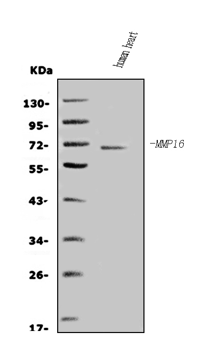
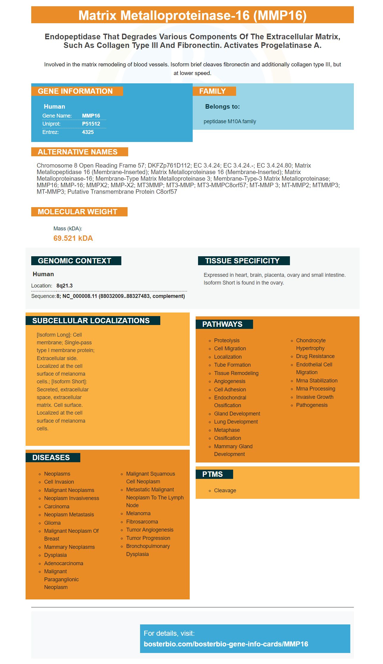
Involved in the matrix remodeling of blood vessels. Isoform brief cleaves fibronectin and additionally collagen type III, but at lower speed.
| Human | |
|---|---|
| Gene Name: | MMP16 |
| Uniprot: | P51512 |
| Entrez: | 4325 |

| Belongs to: |
|---|
| peptidase M10A family |

chromosome 8 open reading frame 57; DKFZp761D112; EC 3.4.24; EC 3.4.24.-; EC 3.4.24.80; matrix metallopeptidase 16 (membrane-inserted); matrix metalloproteinase 16 (membrane-inserted); matrix metalloproteinase-16; Membrane-type matrix metalloproteinase 3; Membrane-type-3 matrix metalloproteinase; MMP16; MMP-16; MMPX2; MMP-X2; MT3MMP; MT3-MMP; MT3-MMPC8orf57; MT-MMP 3; MT-MMP2; MTMMP3; MT-MMP3; Putative transmembrane protein C8orf57
Mass (kDA):
69.521 kDA

| Human | |
|---|---|
| Location: | 8q21.3 |
| Sequence: | 8; NC_000008.11 (88032009..88327483, complement) |
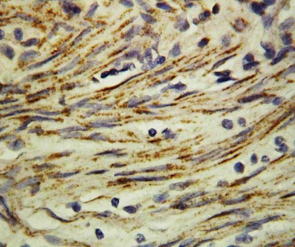
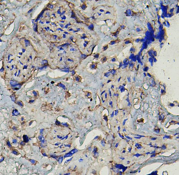
Expressed in heart, brain, placenta, ovary and small intestine. Isoform Short is found in the ovary.
[Isoform Long]: Cell membrane; Single-pass type I membrane protein; Extracellular side. Localized at the cell surface of melanoma cells.; [Isoform Short]: Secreted, extracellular space, extracellular matrix. Cell surface. Localized at the cell surface of melanoma cells.


This is the place to go if you want to learn more about Boster Bio's MMP16 Cell Based ELISA Kit. This article will go over the TCGA cohort, the best cut-off value, as well as how to use it in the clinical setting. We hope these articles will help you in your quest to develop clinical applications of the MMP16 marker.
Boster Bio's MMP16 Cell-based ELIS Kit might be of interest to you if you are looking to test for MMP16 protein phosphorylation. This kit is quick and affordable, and allows researchers to detect quickly changes in the phosphorylation status of targeted cells. It uses a phosphorylation-specific peptide substrate to detect a specific protein.
The enzyme is typically overexpressed in cancer tissues of humans, making it an a crucial tool in tumor research. In animal models, it is used to monitor the development of cancerous cells and their treatment. It is also used in diagnostic procedures. This enzyme can be found in many tissues including the pancreas, the area where it is discovered in the lab. This enzyme is involved in the synthesis of many cellular components, including collagen.
The TCGA PCa methylation data are based on short-term follow-up information and is unable to evaluate the prognostic value of biomarkers that are thought to be methylation-related. The dataset is not representative of the entire spectrum of cancers. Therefore, future studies should be focused on long-term follow-up data. The MMP16 marker is an example of a marker and future studies should investigate its role in predicting the outcome of cancer.
With an TCGA cohort to study the MMP16 gene expression levels in patients suffering from breast cancer. The UALCAN software analyzes these data. This software analyzes the cancer patients' gene sets and provides three survival analysis modules. This application lets users compare gene expression between different types of cancers and look at the impact of SNV frequency and gene methylation.
The TCGA is a major cancer genomics project. In one single study, researchers from 33 different cancer types were matched and molecularly characterised more than 20,000 primary cancer samples. 2.5 petabytes worth of information was generated during this period. It has significantly enhanced the ability to detect and treat cancer. It will be made publicly available for further study. It will also assist doctors in determining the cause of various cancers.
One of the most common questions regarding the identification of an abnormality is "What is the optimal cut-off value for MMP16?" The answer depends on the kind of cancer. Researchers found that patients with melanoma with an MMP-related signature had lower survival rates. They reviewed data from three cohorts made up of 480 patients and calculated a risk score each. The results from the GSE116918 and GSE70769 cohorts revealed poor prognosis for patients that had a MMP-H-related signature. For patients with high levels of MMP16 expression, the HR was 2.54 and AUC for recurrence was 0.711 and 976.
The RNA from these tissues was reverse-transcribed by Takara PrimeScript(r) and kit for RT-PCR (RR036A). The RQ value was used to calculate the relative gene expression levels. The primers used in the analysis included human MMP16S GAPDH F, GAPDH S, and MMP16 R. The RQ value for each gene was calculated with the aid of a computer and an optical microscope.
MMP16 is expressed by tumors with high levels of MMP protein. Patients suffering from various kinds of cancers suffer from a poor prognosis and distant metastasis. MMP16 overexpression in glioma and gastric cancers can cause tumor enlargement and metastasis. A high level of MMP16 expression was also associated with a lower overall survival (OS). Unfortunately, the research does not provide the clinical and molecular information required to establish a high-quality cut-off for MMP16.
The MMP16 marker was initially studied in a clinical trial for gastrointestinal cancer in the USA. However, in the longer future, it could be used to predict the risk of progression to cancer in other tumor types. The genetic risk profile of a patient is used to determine the best cut-off for MMP16 expression. The most appropriate cut-off value for MMP16 expression was determined by normalizing the levels of the sample against GAPDH.
In a paired case-control study MMP16 was detected in tumor tissues. The tumor tissues showed a significantly higher level of MMP16 than normal tissue. This suggests that MMP16 might be an oncogene in HCC. The results showed that MMP16 levels are highly associated with the possibility of developing HCC. Additionally, advanced stages of T and N were associated with better outcomes in patients with high MMP16 levels.
Overexpression of MMPs is a hallmark of the malignant phenotype. Alongside this, increased MMP expression has been observed in the cellular components of primary tumor tissues as well as metastatic tissues. We will review the clinical significance of the MMP16 marker in this article, and the methodological issues to be addressed in order to determine its clinical value. This article reviews some of the current evidence supporting the use of the MMP16 marker in oncology.
MMP16 markers do not provide specificity and therefore are not useful in cancer diagnosis. The 18 members of the MMP16 gene family are structurally related. They are classified into five classes: collagenases gelatinases, stromelysins and non-classified MMPs. Each MMP type contains an peptide signaling with a propeptide domain, zinc binding site, and a haemopexin-like domain.
MMP16 is used to evaluate MMP activity in patients with chronic diseases such as rheumatoid or osteoarthritis. This enzyme is responsible for the destruction of extracellular matrix, and it can be blocked to assist sufferers with their conditions. Inflammatory bowel disease is a common cause of MMPs and the rheumatoid arthritis condition is usually due to their overexpression.
MMPs are zinc-dependent endopeptidases that break down virtually every component of the extracellular matrix. Despite their obvious importance in tissue destruction, MMPs also have anti-inflammatory properties, and they can also process anti-inflammatory cytokines. They also play a role in cell signaling and can regulate the release of neoepitopes. As a result, MMPs are a key element in the physiologic functions of cells.
MMPs play a crucial role in bone formation. In one study, a case of multicentric osteolysis was reported. The patient had low levels of pro MMP-2 in their serum and had 2 specific mutations in the MMP2 gene. This case study highlights the many physiological roles played by MMPs. Additionally, MMPs could also play a role in the development of dental caries.
Most MMPs are secreted in latent precursors that are then activated proteolytically outside the cell. Pro-MMPs that are inactive are kept in their inactive state by a zinc atom. The pro-MMP's cysteine residue blocks access to catalytic sites. A partial proteolytic denature severs cysteine residues and catalytic sites, which expose active MMP. MMPs are activated in a sequential fashion in extracellular space. The first MMP activates the subsequent.
PMID: 7559440 by Takino T., et al. Identification of the second membrane-type matrix metalloproteinase (MT-MMP-2) gene from a human placenta cDNA library. MT-MMPs form a unique membrane-type subclass in the MMP family.
PMID: 9396633 by Matsumoto S., et al. Identification of soluble type of membrane-type matrix metalloproteinase-3 formed by alternatively spliced mRNA.
*More publications can be found for each product on its corresponding product page