This website uses cookies to ensure you get the best experience on our website.
- Table of Contents
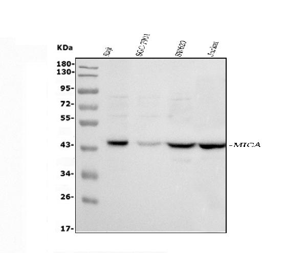
Facts about MHC class I polypeptide-related sequence A.
Ligand for the KLRK1/NKG2D receptor. Binding to KLRK1 leads to cell lysis.
Human | |
---|---|
Gene Name: | MICA |
Uniprot: | Q29983 |
Entrez: | 100507436 |
Belongs to: |
---|
MHC class I family |
FLJ60820; MGC111087; MICA; PERB11.1
Mass (kDA):
42.915 kDA
Human | |
---|---|
Location: | 6p21.33 |
Sequence: | 6; NC_000006.12 (31400711..31415315) |
Widely expressed with the exception of the central nervous system where it is absent. Expressed predominantly in gastric epithelium and also in monocytes, keratinocytes, endothelial cells, fibroblasts and in the outer layer of Hassal's corpuscles within the medulla of normal thymus. In skin, expressed mainly in the keratin layers, basal cells, ducts and follicles. Also expressed in many, but not all, epithelial tumors of lung, breast, kidney, ovary, prostate and colon. In thyomas, overexpressed in cortical and medullar epithelial cells. Tumors expressing MICA display increased levels of gamma delta T-cells.
Cell membrane; Single-pass type I membrane protein. Cytoplasm. Expressed on the cell surface in gastric epithelium, endothelial cells and fibroblasts and in the cytoplasm in keratinocytes and monocytes. Infection with human adenovirus 5 suppresses cell surface expression due to the adenoviral E3-19K protein which causes retention in the endoplasmic reticulum.
This article will describe the flow the cytometry workflow and the benefits of the MICA Marker made by Boster Bio. The advantages of the MICA Marker are discussed as well as the MIC MAbs with different characteristics. The article also discusses the importance of QC, as well as the various types of buffers for lysing. Using the correct buffer for lysing is crucial to getting good results.
The Boster Bio flow cytometry protocols series provides an easy-to-follow overview of the method, including preparation of samples, tips for optimizing and troubleshooting suggestions. The protocols employ the MICA marker, which is a popular biomarker that is used to identify cells within a fluid. FACS, or fluorescence-activated cell sorting, allows researchers to identify and profile cells in fluid mixtures. The binding of antibodies may alter the expression of intracellular signalling proteins by altering their surface antigens. Saponin and Methanol do not alter the epitopes of the surface antigens and are therefore compatible with this marker.
Many Boster Bio flow-cytometry protocols employ MICA to detect cells in tissues and blood. For each test, a table is provided with the stimulants, the incubation times and the target proteins. Staining is available using brefeldin A or monensin. Aliquots that are not stained are used as the baseline control. Visit the Boster Bio website for more information.
The MICA marker is the preferred option for flow cytometry because it is extremely specific in the detection of an antigen. This marker is widely used in immunotherapy for cancer, such as Ovarian Cancer. Boster Bio's flow-cytometry protocols use MICA to identify cancer cells in different tissues. They also offer several other protocols that have been proven to be effective.
Human cells express hundreds of thousands of cell surface antigens, and flow cytometry uses fluorochrome-conjugated antibodies to analyze these proteins directly. This method eliminates the risk of non-specific binding that is possible with indirect staining using immunofluorescence. However, this method does require additional staining using secondary antibodies. The MICA marker is distinct from indirect immunofluorescence staining in that the primary antibody-fluorophore conjugate is directly conjugated with the fluorophore directly. This eliminates the possibility of non-specific binding resulting from large antibody-fluorophore complexes. Additionally, secondary antibodies may not be able to get to cells.
The MILC reagent contains an DILC mimic that is synthesized by Guangzhou RiboBio Co., Ltd. and used to transfect THP-1 cells. It contains 10mM FBS 100 mg/ml streptomycin Sulfate, and 100 U/ml penicillin sodium. The cells were kept in a humidified environment with 5 percent CO2 at 37 degrees Celsius. The TaqMan micro (mi-)RNA reverse transcription kit was used to reverse-transcribe the RNA to DNA. To reverse-transcribe RNA, primers for STAT3, TLR4, DILC and DILC were used.
Flow cytometry data analysis workflows usually comprise worksheets that include a series of plots, each which displays the signal intensity of one or more markers. Every event in an experiment is plotted and displayed in various ways. To determine the number of people of the interest, there are numerous data plots and gates. These are based on gating hierarchy or boolean logic. After that, the data can be analysed by calculating statistics on each data point within a gate.
When analyzing cell populations, flow analysis should be repeated. Multiple peaks can be seen on the histogram if flow analysis is conducted on cells that have mixed populations. A negative isotype control must be carried out on the positive data in such cases. If there is a significant change in intensity between the negative control and the positive samples, this suggests that the cell population is dominated by MICA-positive cells.
The use of a cluster of differentiation markers allows you to precisely identify leukocyte subsets. This marker is essential for the consistent identification and subsets of leukocytes. While quantitative information about CD markers isn't available yet however, the workflow is a useful tool to determine the number of cells and to determine their characteristics. By using a MICA marker, researchers can compare expression patterns across samples, thereby defining new clusters of differentiation.
The preparation of samples is crucial for flow cytometry to work properly. Single particles are prepared in single particle suspensions. They are then passed through a laser beam to analyze the signal. The resultant data is sorting and counting. A well-planned sample preparation process is essential to the success of your research. Overly large amounts of debris, for example could increase noise and make it difficult to locate the cells of interest.
Fluorescently conjugated antibodies have been an effective tool in flow analysis using cytometry. These antibodies identify specific cells with specific structures. They create a pulse of photon emission when they interact with a laser beam. These pulses are detected by a charge-coupled devices, called PMTs. The intensity of fluorescence is directly proportional to the area of the voltage pulse. Therefore, the greater intensity of fluorescence, the more channels there are.
By staining paraffin embedded tissue stained with formalin with mAbs for MICA or its denatured forms, we tested the specificity of MIC MAbs. All mouse immune sera were positive for MICA positive cells, however some hybridomas were positive for one specific method. The various MIC mAbs had different specificities for MICA, MICB, or both.
The soluble form MIC was detected in sera of cancer patients. It was found to be associated with impairment of NKG2D dependent activation of immune cells, which leads to a reversal of immune defense. Numerous studies on immunotherapy and cancer gene therapy have been conducted on the basis of the recognition MICA/NKG2D. These studies have proven that there is a high chance of clinical success. While more research is required to verify these results, the mAbs already show promising potential as therapeutic agents.
LST-007 is a specific target within the whole PA bacteria. However, its recognition of flagellin type B is blocked by glycosylation the molecule. This was the motivation for further bioactivity tests. The LST-007 mAb was capable of binding PA bacteria that were intact. The human isotype control antibody mAb was absent, which led to lower reactivity.
Incredibly, the anti-MICA mAbs WW2G8, WW6B7, and WW9B8 showed distinct characteristics. All three were reactive to the 62 kDa MICA proteins in BL21 cell-lysate as well as 38kDa MICA proteins in HeLa cells. However, WW2G8 and WW6B7 reacted poorly with the native form of MICA on HeLa cells, suggesting they may not be particularly sensitive against the denatured form.
PMID: 8022771 by Bahram S., et al. A second lineage of mammalian major histocompatibility complex class I genes.
PMID: 8613147 by Bahram S., et al. Nucleotide sequence of the human MHC class I MICA gene.