This website uses cookies to ensure you get the best experience on our website.
- Table of Contents
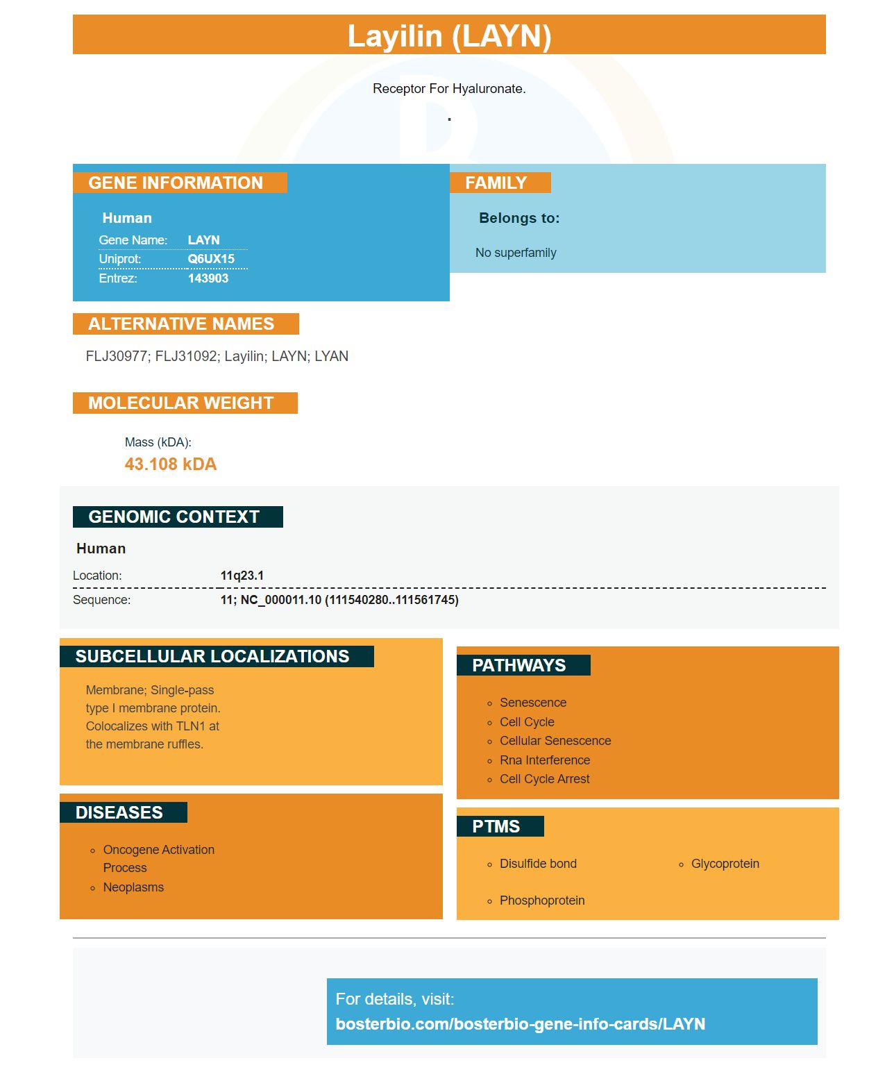

Facts about Layilin.

Receptor for hyaluronate.
.| Human | |
|---|---|
| Gene Name: | LAYN |
| Uniprot: | Q6UX15 |
| Entrez: | 143903 |

| Belongs to: |
|---|
| No superfamily |

FLJ30977; FLJ31092; Layilin; LAYN; LYAN
Mass (kDA):
43.108 kDA

| Human | |
|---|---|
| Location: | 11q23.1 |
| Sequence: | 11; NC_000011.10 (111540280..111561745) |
Membrane; Single-pass type I membrane protein. Colocalizes with TLN1 at the membrane ruffles.


The LAYN protein is a fluorescent protein that binds specifically to the LAYN genetic code. This enzyme has many applications, including the study gene expression and tumor development. ECL and DAB methods for protein transfer are excellent options. Boster Bio is pleased that it offers high-affinity primaries antibodies.
A variety of proteins can be used to detect and analyze LAYN protein expression in live cells. Boster Bio has produced high-affinity primary antibodies against the LAYN marker for its customers. This product line includes 38 protein antibody that are sensitive to this marker as well as 22 immunocytochemistry-grade antibodies. The antibody repertoire contains proteins that are involved with membrane trafficking, perception and hormone synthesis.
The company created a proprietary extraction procedure to obtain high-affinity primaries antibodies against the LYN marker. First, 150 mls antigen were diluted with 50 ul of 1M Tris-HCl. Then, 100 mM of Glycine was added to the mixture. The secondary antibody was then added. The sample was then rinsed twice with TBST.
Enhanced chemiluminescence, a chemiluminescence detection technique, uses an antibody conjugated in HP. When the conjugation of an antibody is applied to a material, it creates excited intermediates that emit light with a wavelength of 450 nm. These excited intermediates will be released during the enzyme/substrate interaction. The signal output stops if the substrate has no ligand.
The light emissions produced are captured with an x-ray film. An instrument that uses luminescent signals provides greater sensitivity, resolution, and dynamic range. In some cases, researchers may be able to manipulate images in real time using cooled CCD cameras. Most researchers still prefer to capture data using film. The light emission creates a dynamic signal, which fades exponentially over time.
Both the ABC- and DAB colorimetric measurement methods use the exact same principle to measure the concentration of proteins. But the Boster ECL chemiluminescent systems uses a different detection system that is based upon the substances bound the secondary antibody. This method uses an enhancer which is based on exposure of the secondary antigen to a substrate. It's faster than the ABC-colorimetric detection method. The DAB enhancer is used by Abcam to validate antibodies.
DAB staining involves adding a small amount of hydrogen peroxide to the HRP-stained sections. These sections are then incubated for several minutes in the DAB solution and then rinsed with a buffer and water series. The sections are then counterstained, dried in ethanol series, and mounted. The DAB staining is often monitored by a microscope. It is recommended to stop the procedure when the section appears light brown in color.
DAB methods can be used to detect neurodegenerative diseases biomarkers in foods. These methods use nanoparticles made from bioactive molecules, such as dopamine, to detect drugs in a variety o biological samples. The results are reliable and have been highly cited in 104 papers. These methods can also be used to identify biomarkers within human urine.
Membrane stining is an essential step in protein transfusion experiments. You need to use the right staining solutions to achieve high-quality membrane pictures. This buffer contains 25 mg Tris base, 190 mg glycine and 20% methanol. Once the gel is prepared, it should be placed between filters containing transfer buffer. The sandwich should be left in the buffer for at most 15 minutes. Multiple exposures will be required to ensure a high signal–to–noise ratio.
To increase the sensitivity of ELISA kits, Boster Bio developed Picokine(tm). This platform is capable of enhancing sensitivity to the picogram level. Picokine antibodies have been tested against a variety sample types. On request, validation images and procedures can be obtained. Supervision, a secondary antibody made of polymer-based polymers, can reduce IHC time up to 30%. The technology is supported by insights into immunogen design and BeNeLux distributor Sanbio.
The western blotting process requires pooled serum proteins from humans to be separated with 10% SDS/PAGE. The separated proteins then go through a Tris-glycine, SDS transfer buffer to transfer them to PVDF or nitrocellulose cells. For fast and reversible detections, the membranes are stained with Coomassie Brilliant Blue R250. The method also allows for a rapid and reliable detection of protein bands with a high level of reproducibility.
Membrane-based selection allows researchers to choose a more efficient membrane staining method based on the target proteins. When membrane proteins are purified, they often lose their natural structures. Boster Bio can increase protein transfer efficiency through membrane staining. This method is more effective than the traditional process of using a purified recombinant protein.
PMID: 11294894 by Bono P., et al. Layilin, a novel integral membrane protein, is a hyaluronan receptor.


