This website uses cookies to ensure you get the best experience on our website.
- Table of Contents
1 Citations 6 Q&As
1 Citations 7 Q&As
Facts about Lymphatic vessel endothelial hyaluronic acid receptor 1.
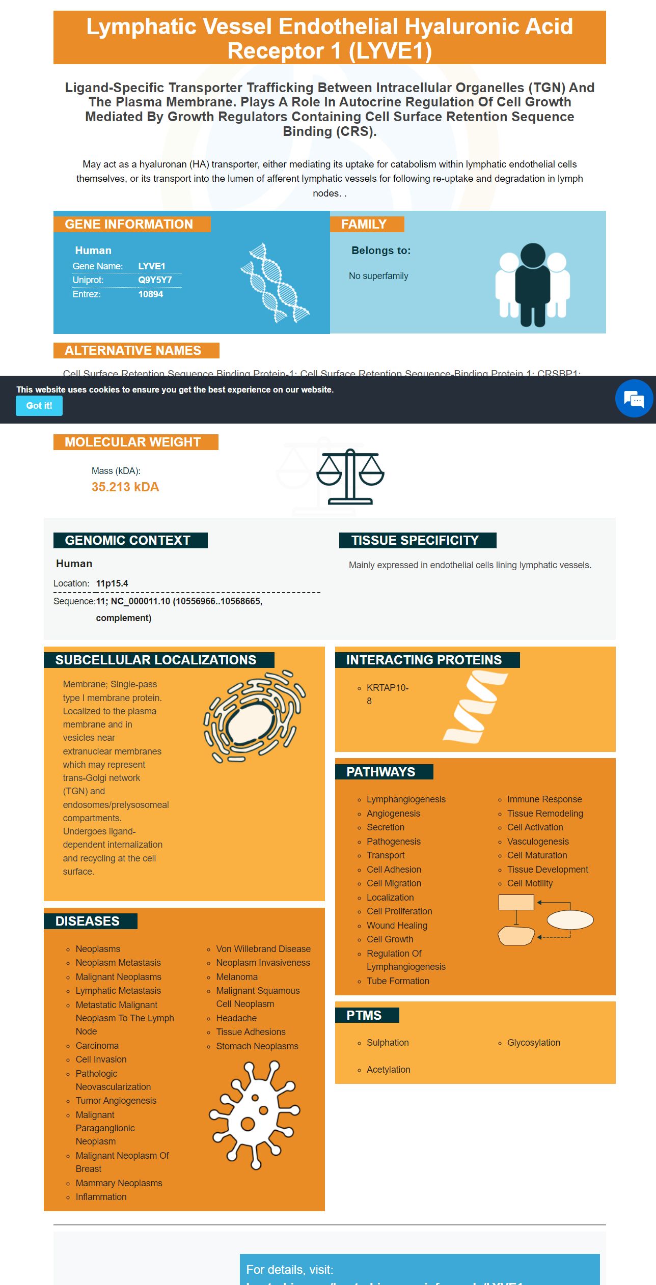
May act as a hyaluronan (HA) transporter, either mediating its uptake for catabolism within lymphatic endothelial cells themselves, or its transport into the lumen of afferent lymphatic vessels for following re-uptake and degradation in lymph nodes. .
Human | |
---|---|
Gene Name: | LYVE1 |
Uniprot: | Q9Y5Y7 |
Entrez: | 10894 |
Belongs to: |
---|
No superfamily |
cell surface retention sequence binding protein-1; Cell surface retention sequence-binding protein 1; CRSBP1; CRSBP-1; extracellular link domain containing 1; extracellular link domain-containing 1; Extracellular link domain-containing protein 1; HAR; Hyaluronic acid receptor; lymphatic vessel endothelial hyaluronan receptor 1; lymphatic vessel endothelial hyaluronic acid receptor 1; LYVE1; LYVE-1; LYVE-1XLKD1; XLKD1
Mass (kDA):
35.213 kDA
Human | |
---|---|
Location: | 11p15.4 |
Sequence: | 11; NC_000011.10 (10556966..10568665, complement) |
Mainly expressed in endothelial cells lining lymphatic vessels.
Membrane; Single-pass type I membrane protein. Localized to the plasma membrane and in vesicles near extranuclear membranes which may represent trans-Golgi network (TGN) and endosomes/prelysosomeal compartments. Undergoes ligand-dependent internalization and recycling at the cell surface.
The LYVE1 marker is an integral membrane glycoprotein of type I that is expressed on the lymphatic endothelium. It is a receptor to the mucopolysaccharide (hyaluronan) Continue reading to learn more about its best uses, as well as how it can be used in diagnosis and research. This article also covers the LYVE1 antibody.
LYVE-1, a homolog to CD44, is a cell surface glycoprotein required for HA anchoring onto vascular endothelial tissues. It is still not clear what LYVE-1 does to bind HA. However, it has been associated with HA transport and turnover and has been shown to promote HA localization on the surface of lymphatic endothelium. Numerous studies have also shown LYVE-1's utility in lymphoid tissues and lymphangiogenesis.
LYVE-1 belongs to the Link protein family and is characterized as a consensus hyaluronan–binding domain. Although LYVE-1 may have many different regions, it shares many similarities to CD44. Among them is the conserved hyaluronan binding domain in the extracellular domain. Additionally, LYVE-1 is part of the lymphatic system, which transports proteins and cells to organs and tissues and serves as an immune surveyor. Hyaluronan, an extracellular matrix mucopolysaccharide can bind with LYVE-1 in vivo.
Peripheral tissues have low levels of hyaluronan. Their levels can rise in response o lymphedema and infection. A disruption in lymph flow can also alter the rate of hydrochloric acid catabolism. Lymphatics play an essential role in hyaluronan Homeostasis.
LYVE1 binds hyaluronic and is a protein. It is located on the surface of lymphatic endothelial cells and may have multiple functions. LYVE1 is a marker of lymphatic endothelial cell function and plays a role in lymphatic transport. However, the physiological role of LYVE1 is not clear at present. Its exact function will require more research.
This gene encodes a type 1 integral membrane glycoprotein. LYVE1 can be found in the lymphatic endothelium. This gene is thought to be involved in regulating traffic between tumor cells and lymph nodes. Lymphatic vessels are a secondary circulatory system that drains extracellular liquid from tissues and provides an environment for immune cells, which can encounter antigen. Lymphatic endothelium contains markers such as LYVE1, PALE, VEGFR3, podoplanin, and VEGFR3 (the receptor for VEGF).
LYVE1 plays a role in transporting HA, but it also mediates catabolism of lymphatic endothelial tissues. LYVE1 delivers HA to lymphatic vessels for reuptake by lymph nodes. The lymphatic system can degrade HA in lymph nodes and fragments may migrate into the blood. Once HA fragments have reached the bloodstream they are broken down and transported to liver.
LYVE1 binds HA (a component of extracellular matrix) via a transmembrane. HA is abundantly found in connective tissues and skin. LYVE1 is most commonly found in lymphatic vessels lining endothelial cells. Its half-life spans 24 hours, and it is classified a type 1 integral membrane glycoprotein. Lyve1 can both bind soluble and immobilised human HA.
LYVE1 can also be known as lymphatic vessel receptor-1. It is a member of the link proteins family, which binds to hyaluronan. It is found in lymphatic endothelial tissues and may play an important role in tumor metastasis. Its functions are not yet understood, but it is an important marker for lymphatic endothelial cells.
LYVE1 was identified as a crucial surface marker for lymphatic vessels. Although there are no obvious lymphatic defects caused by LYVE-1 deletion, high levels of LYVE-1 can have profound effects on lymphangiogenesis. Recent research has shown that CD44, an important regulator of developmental processes such as metastasis and the progression of cancer, is elevated in many cases. Therefore, LYVE1 may be a useful molecular marker for understanding lymphangiogenesis.
It is unusual for LYVE1 to be expressed on the lymphatic epithelium. This protein isn't found in any other tissues, such as epithelial and fibroblast cells. Contrary to CD44 which is found in multiple sizes and isoforms of CD44, LYVE-1 appears only to be expressed as a single species. Additionally, immunohistochemistry has detected the protein using specific antibodies.
During embryogenesis, LYVE1 was expressed on lymphatic vessels and major venous blood vessels. LYVE-1 expression can be detected in CD31+ endothelial tissues of the lung, liver, and other organs. However, this expression is not maintained and is lost upon vessel development. Lymphatic endothelia were, however, positively stained by Prox1 antibody. This antibody excludes other tissues, such as the primary sinus and intersegmental.
The role played by LYVE-1 in HA transportation is not known. The receptor has been implicated for the catabolism hyaluronan within the lymph nodes (liver, and spleen). Lyve-1, a novel receptor for Hyaluronan, is necessary to compensate CD44's loss. Therefore, LYVE-1 is an important marker in the study of lymphangiogenesis and lymphoid tissue.
Boster Bio has validated the Anti-CD31/PECAM-1 Monoclonal Antibody for use in IHC-P. This antibody binds to human and rabbit forms of this specific protein. It has a high affinity and has been extensively quoted over the last 25-years. Boster antibodies have also undergone multiple validations in the immunohistochemistry, Western blotting, and ELISA fields.
There are two types basic ELISAs: qualitative and quantitative. A quantitative ELISA is a method that measures the concentration of analyte and returns a simple positive/negative result. A qualitative ELISA determines whether analyte is present or absent. A quantitative ELISA measures analyte concentration using a standard graph. Both quantitative and qualitative ELISAs are equally effective. However, the latter is more commonly used for clinical purposes.
ELISA is an immunoassay procedure that measures a protein’s concentration by binding to antibodies. It is also used to determine the level of reproductive hormones in various species and to evaluate them within the context of conservation efforts. The ELISA kits are available in a variety of formats. There are three options: a complete kit, preoptimized and antibody pairs, or a pre-optimized reagents. There are ELISA kits for human, mouse, porcine, bovine, and equine species, as well as custom targets.
A high quality ELISA kit can provide reliable and relevant results to your research. Its performance is measured by parameters like linearity of dilution, recovery, and precision. These are important ELISA parameters as they will help you determine if your samples work well with the kit. A competitive ELISA test kit will give you reproducible results. Enzo Life Sciences can provide high-quality ELISA Kits at affordable prices.
Multiple samples of serum and tissue have been used to validate the Boster Bio LYVE1 marker. The LYVE1 protein is found in macrophages as well as dendritic cell dendritic cells. It is involved in lymph vessel entry and migration. This antibody detects both populations. It has high specificity and sensitivity, making it ideal for a variety sample types.
The tumor tissue was immunostained with antibodies against BrdUrd or AdVEGFR-3Ig to show the LYVE1 gene. LYVE-1 staining revealed lymphatic vessel sprouting in tumor tissues. Lymphatic vessels could be found in tumor tissues at week two and three, but they were absent at the beginning of week one. Lymphatic vessels were also found around tumor cells when cocultured vitro.
Boster Bio LYVE1Marker is able to detect LYVE1 and was validated with a variety of samples. It was used for analyzing the expression of a variety of endothelial marker in human tissues. This antibody is also compatible with a variety different sample types. The BosterBio LYVE1 marker has been successfully validated in many samples. It can be used for differentiation between different cell types.
PMID: 10037799 by Banerji S., et al. LYVE-1, a new homologue of the CD44 glycoprotein is a lymph-specific receptor for hyaluronan.
*More publications can be found for each product on its corresponding product page