This website uses cookies to ensure you get the best experience on our website.
- Table of Contents
1 Citations
Facts about ATP-sensitive inward rectifier potassium channel 8.
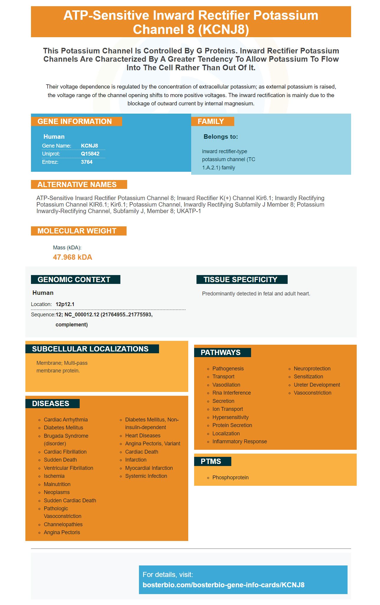
Their voltage dependence is regulated by the concentration of extracellular potassium; as external potassium is raised, the voltage range of the channel opening shifts to more positive voltages. The inward rectification is mainly due to the blockage of outward current by internal magnesium.
Human | |
---|---|
Gene Name: | KCNJ8 |
Uniprot: | Q15842 |
Entrez: | 3764 |
Belongs to: |
---|
inward rectifier-type potassium channel (TC 1.A.2.1) family |
ATP-sensitive inward rectifier potassium channel 8; Inward rectifier K(+) channel Kir6.1; inwardly rectifying potassium channel KIR6.1; Kir6.1; Potassium channel, inwardly rectifying subfamily J member 8; potassium inwardly-rectifying channel, subfamily J, member 8; uKATP-1
Mass (kDA):
47.968 kDA
Human | |
---|---|
Location: | 12p12.1 |
Sequence: | 12; NC_000012.12 (21764955..21775593, complement) |
Predominantly detected in fetal and adult heart.
Membrane; Multi-pass membrane protein.
There are a variety of reasons to purchase Boster Bio's Anti-Kir6.1/KCNJ8 Marker. From Steven Boster's gene infographics to Boster's validation on immunohistochemistry and Western Blotting, we have you covered. Read on to learn more about this new product from Boster Bio. This product is suitable for researchers worldwide.
The KCNJ8 gene encodes a protein called potassium inwardly-rectifying channel subfamily J. This protein is a component of the membrane of most mammalian cells and is involved in several physiologic functions. The protein is controlled by G-proteins, and defects in this gene result in a variety of symptoms including J-wave syndromes and sudden infant death syndrome.
In the validation of primary antibodies on Western Blotting, Boster's scientists look for two primary requirements: specificity and affinity. The latter requires that the antibody recognize its target protein in the assay for which it is designed. Detection of new bands is a signal that the assay needs to be optimized or that the primary antibody must be changed. A control blot is a helpful tool in identifying potential cross-reactivity. Secondary antibodies may be conjugated to different enzymes or fluorophores to distinguish them from one another.
To validate the specificity of primary antibodies on Western Blotting, the antigens are mixed in two separate cell lines or tissues. These samples are compared with each other, and the signals from each cell line are analyzed by using a CCD-camera system. In addition, the results of Western blotting are compared to the RNA-Seq data of the same samples. To ensure that antibodies are specific, positive controls are used to assess the results. Positive controls are a good way to check whether an antibody is specific for a given target protein. The antigens used in orthogonal validation are cell lines or tissues with a five-fold difference in expression.
Detailed information on the validity of primary antibodies is necessary for peer-review. This information should include the characteristics of the primary antibodies tested and the results should be consistent. The raw data should include information on dilutions, incubation times, positive and negative controls, sample information, and organism/species specificity. These are just a few of the characteristics that should be included in the validation of primary antibodies on Western Blotting.
KO validation of antibodies is considered the gold standard for Western blotting and is often used by antibody vendors during batch testing. Assay-specific validation requires a combination of KO and nonspecific validation. In addition, the antibody supplier/distributor can perform assay-specific validation for its products. The following are the two strategies used in assay-specific antibody validation. There are many other approaches to validate primary antibodies on Western blotting.
In the field of immunohistochemistry, a validation process is required for primary antibodies. This process involves the testing of the specificity of the antibodies against various targets. This is a complex and laborious process, and may not be required for antibodies with known distribution patterns. An informal survey was conducted to collect opinions on the subject of antibody validation. The aim of the survey was to initiate a conversation regarding the requirements of Frontiers journal.
One of the most important criteria for validation is the ability to recognize the positive and negative controls on the tissue. While positive controls must express the target protein, a negative control sample should not. A positive tissue is a useful way to determine the correct cell type and compartment of staining. Positive tissue samples should also contain matched normal tissues. To validate an antibody, it should also be tested on tissue of a variety of pathologies and cell lines.
The second important factor for antibody validation is RNA consistency. This is evaluated using the staining pattern of two primary antibodies that do not have any overlap in epitopes. In this process, two antibodies must show similar staining patterns in at least two tissue samples or cell lines. Additionally, a validation process can involve over-expression of the target protein in the cell line to determine if the antibody is able to detect it on a cellular level.
Another aspect of antibody validation that is frequently ignored is the antigen recognition. In western blot detection, the antigen is an amino acid stretch after denaturing SDS-PAGE. In immunoisolation and immunoprecipitation, the antigen has a more native or cross-linked structure. As a result, the antibodies may fail to detect the target if they have different properties in these different applications. Therefore, validation of primary antibodies is needed to assess their performance across multiple applications.
In addition to ELISA testing, the Boster's primary antibodies are also validated against the target protein using siRNA transfection. The technique involves spleen cells that respond to the antibody. Then, the best responding animals are selected for hybridoma fusion. The supernatants from the best-responding animals undergo an ELISA assay. The selected antibodies undergo further application testing before they are released into the market.
PMID: 8595887 by Inagaki N., et al. cDNA sequence, gene structure, and chromosomal localization of the human ATP-sensitive potassium channel, uKATP-1, gene (KCNJ8).
PMID: 9573340 by Erginel-Unaltuna N., et al. Genomic organization and expression of KCNJ8/Kir6.1, a gene encoding a subunit of an ATP-sensitive potassium channel.
*More publications can be found for each product on its corresponding product page