This website uses cookies to ensure you get the best experience on our website.
- Table of Contents
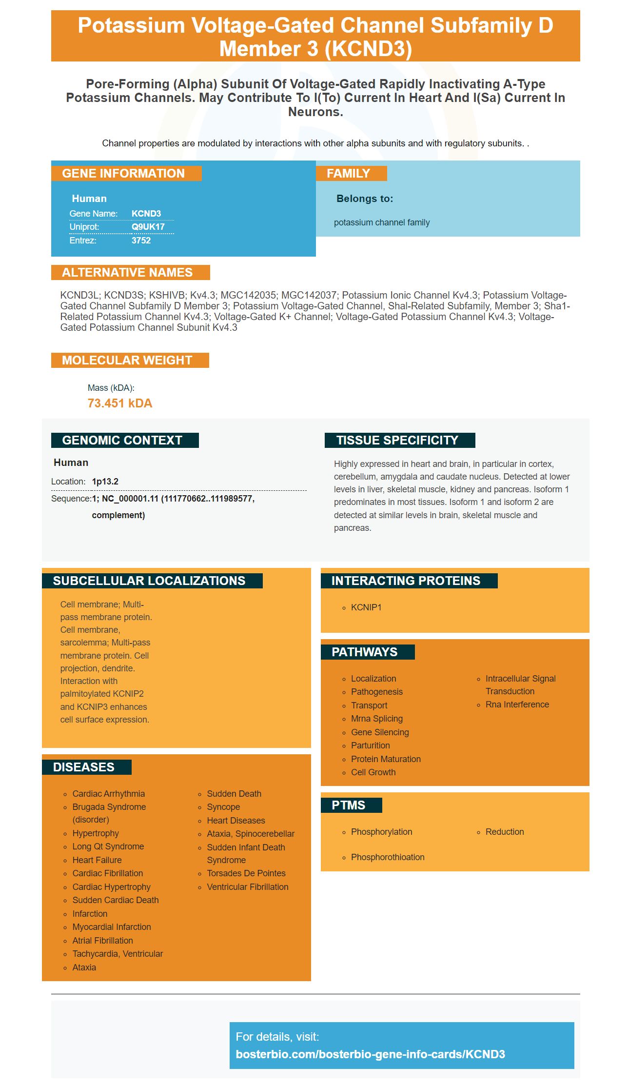
Facts about Potassium voltage-gated channel subfamily D member 3.
Channel properties are modulated by interactions with other alpha subunits and with regulatory subunits. .
Human | |
---|---|
Gene Name: | KCND3 |
Uniprot: | Q9UK17 |
Entrez: | 3752 |
Belongs to: |
---|
potassium channel family |
KCND3L; KCND3S; KSHIVB; Kv4.3; MGC142035; MGC142037; potassium ionic channel Kv4.3; potassium voltage-gated channel subfamily D member 3; potassium voltage-gated channel, Shal-related subfamily, member 3; sha1-related potassium channel Kv4.3; voltage-gated K+ channel; voltage-gated potassium channel Kv4.3; Voltage-gated potassium channel subunit Kv4.3
Mass (kDA):
73.451 kDA
Human | |
---|---|
Location: | 1p13.2 |
Sequence: | 1; NC_000001.11 (111770662..111989577, complement) |
Highly expressed in heart and brain, in particular in cortex, cerebellum, amygdala and caudate nucleus. Detected at lower levels in liver, skeletal muscle, kidney and pancreas. Isoform 1 predominates in most tissues. Isoform 1 and isoform 2 are detected at similar levels in brain, skeletal muscle and pancreas.
Cell membrane; Multi-pass membrane protein. Cell membrane, sarcolemma; Multi-pass membrane protein. Cell projection, dendrite. Interaction with palmitoylated KCNIP2 and KCNIP3 enhances cell surface expression.
This page contains information about antibodies to KCND3. Here you will discover the benefits of using high-affinity antibodies to the KCND3 marker. These antibodies can greatly increase the success of your experiment. Read on for more information! Listed below are some of the best uses for the KCND3 Marker. Catalog number A03317-1 refers to the boster Bio anti-KCND3 Antibody. The bosterbio Anti-KCND3 antibody reacts against the human, mouse and rat proteins.
Boster Bio Anti KCND3 Marker reacts with Human, Mouse, Rat, and Rat. Its catalogue number is A03317-1. There are many other KCND3 antibodies. Boster Bio's Kv4.3 pore forming antibody anti-KCND3 is one of these. The antibody's primary function is to detect KCND3 cells.
The KCND3 marker was chosen as a good candidate for detecting epitopes in fixed tissue. The KCND3 marker is highly specific for the KCND3 peptide. High affinity is not always desirable. The antibody may be too sensitive to separate the antigen from it, which could require harsh or denatured methods to get rid of it.
Lacking IgD in mice led to delayed generation of high-affinity primary antibody, which resulted in prolonged autoimmune diabetes. IgD plays a crucial role in the transition from primary autoreactive response to secondary antigen–specific antibody responses. The generation of protective memory IgM can be delayed if IgD is absent. Understanding the role of IgD is essential in limiting autoantibody response is important.
We identified IgD, IgG and KCND3 markers. IgD inhibits the release of autoreactive IgG antibody and controls B cell proliferation and activation. In addition, the antibody secreting cells lack typical B1-B cell markers. These results indicate that IgD insufficient B cells are vulnerable for rapid induction of primary immune response.
The IgM class BCR binds to a molecule called hen-egg lysozyme, which induces a calcium flux in IgM and IgD B cells. Complex HEL blocked the activation IgD-deficient IgD B cells. The flexible hinge region of the BCR is the cause of this partial polyreactivity. It is also thought that B-lymphocytes lacking IgD have a lower threshold for activation.
The KCND3 marker was also used for high-affinity antibodies. These antibodies are highly specific for IgG/IgM. They are also effective in producing monoclonal, high-affinity monoclonal antibodies. This research has provided new insights into the production monovalent antibodies. Improved vaccine design, production and manufacturing will be possible by better understanding the mechanism for NP binding in the immune system mouse.
InsAKLH peptide injections were used to inject IgD-deficient mice. The serum of IgD deficient mice was collected on days 0, 21 and 42. The serum immunoglobulin titers were determined by ELISA and were compared to WT controls. All experiments were repeated at least two times to confirm the results.
The KCND3 gene encodes the potassium channel transporter I(tO). Exon 3 of KCND3 contains the T377M mutation. Three independent programs predicted this mutation to be pathogenic. However, the ExAC database and Swedgene do not contain this mutation. Swedgene contains genome data on approximately 1000 human subjects. This mutation is classified as a Class 5 variant by ACMHG. This marker can be used for screening for disease-causing variants.
Four mutations of the KCND3 gene were identified in Brugada Syndrome. These mutations were found at S6 transmembrane Domain A. This marker has a non-clear genotype-phenotype correlation. However, there is strong evidence for risk stratification in patients suffering from KCND3 mutations. To confirm the results, and to determine the role of KCND3 within this syndrome, additional neuropathological research is needed.
Multiple studies have shown that KCND3 variants are associated cardiac arrhythmias. Brugada Syndrome and other related cardiocerebral problems are known to be caused by this gene. KCND3 is widely expressed in the brain, with higher expression in the heart and cerebellum. Cardiomyocytes express KCND3 within the heart, which is critical for action potential repolarization.
In a large UK cohort of ataxia patients, three patients with SCA19/SCA22 were identified. Similar results were obtained when screening was done in a smaller Asian population. It has been proven that KCND3 mutants associated with ataxia is highly penetrant. The Swedish family also has a high penetrance for these mutations. Only one KCND3 mutation has been identified as having a reduced penetrance in this disease.
PMID: 9843794 by Kong W., et al. Isolation and characterization of the human gene encoding Ito: further diversity by alternative mRNA splicing.
PMID: 10200233 by Dilks D., et al. Cloning and expression of the human Kv4.3 potassium channel.