This website uses cookies to ensure you get the best experience on our website.
- Table of Contents
Facts about Interleukin-1 receptor-associated kinase 1.
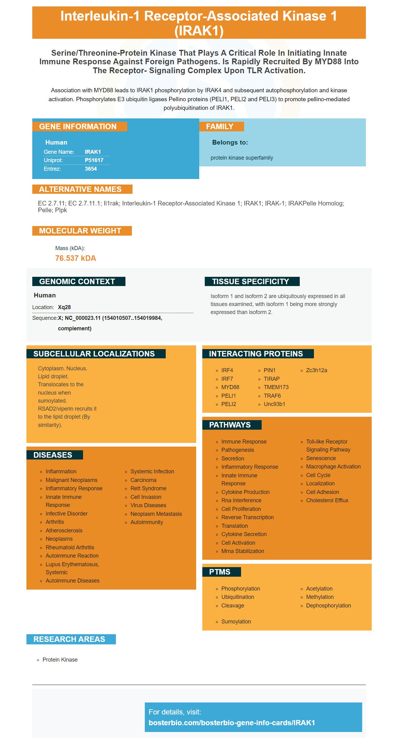
Association with MYD88 leads to IRAK1 phosphorylation by IRAK4 and subsequent autophosphorylation and kinase activation. Phosphorylates E3 ubiquitin ligases Pellino proteins (PELI1, PELI2 and PELI3) to promote pellino-mediated polyubiquitination of IRAK1.
Human | |
---|---|
Gene Name: | IRAK1 |
Uniprot: | P51617 |
Entrez: | 3654 |
Belongs to: |
---|
protein kinase superfamily |
EC 2.7.11; EC 2.7.11.1; Il1rak; interleukin-1 receptor-associated kinase 1; IRAK1; IRAK-1; IRAKPelle homolog; Pelle; Plpk
Mass (kDA):
76.537 kDA
Human | |
---|---|
Location: | Xq28 |
Sequence: | X; NC_000023.11 (154010507..154019984, complement) |
Isoform 1 and isoform 2 are ubiquitously expressed in all tissues examined, with isoform 1 being more strongly expressed than isoform 2.
Cytoplasm. Nucleus. Lipid droplet. Translocates to the nucleus when sumoylated. RSAD2/viperin recruits it to the lipid droplet (By similarity).
The IRAK1 marker may be a target for miR204. It has been established that miR-204 is able to target various proteins, including IRAK1. The IRAK1 protein is located in the nucleus of numerous cells and may be useful for a variety of biological applications. In this article, we'll examine its potential applications in vivo and in vitro. We will also explore its potential for future use in other biological applications.
MiR-204 block the activity of a tumor suppressor , known as VHL. VHL is recognized for its ability to restore miR-204 activity in tumors, including esophageal squamous cell carcinoma. Downregulation of miR-204 inhibits autophagy and enhances Caspase-3/9-dependent apoptosis in cancer cells. MiR-204 could be a treatment target for GC.
MicroRNAs are vital non-coding RNAs that regulate about 30% of the human genome. They bind with specific regions of the 3'UTR of target molecules to block their activity. Different mechanisms are employed to induce silencing of mRNAs through miRNAs. These mechanisms are dependent on the mutual complementarity between miRNA seed sequences and the mRNA target. Apart from targeting the IRAK1 genes, miR204 regulates many other genes, such as the RNA kinases and RNA polymerase II.
Numerous studies have revealed that autophagy is controlled by miRNAs. One study showed that miR204 reduces autophagy-related CUP5 proteins in Caenorhabditis elegans, and inhibits the function IRAK1 in primary cortical neurons in humans. Many aging processes have been related to miRNAs which regulate autophagy in humans. Autophagy levels that are low have been linked to intervertebral disk degeneration.
MiR-146a and IRAK1 are two other possible miR-204 targets. Both miRs have a 2-nt difference at their 3' ends and could function as innovative negative regulators of the immune response. They may also aid in fine-tuning the immune response by controlling TRAF6 and IRAK1.
MiR-204 is associated with ovarian cancer. It suppresses IGFBP-3 expression, which could play a part in the development of ovarian cancer. It's also associated with an increase in tumor invasiveness, migratory aptitude, and bone metastasis. MiR-146b-5p on the other side, inhibits IL-6 and IL-8. Among miRNAs that regulate NF-kB, miR-30a-5p and miR-18a/b are the three most important.
IRAK1 is an NF-kB-linked gene and is thought to be associated with aggressiveness of disease. DLBCL confirms that a lower number of copies of IRAK1 correlates with a worse prognosis. The phase 1 study of pacritinib in this cancer revealed partial responses in 9 patients with FL or relapsed/refractory NHL.
MiR-204 blocks the development and proliferation of microglial cells both in vitro and in mice. It reduces cerebral infarction volume, inhibits neuroinflammation and inhibits inflammation reactions. In addition, it modulates the expression of SIRT1 and IRAK1.
MiR-204 has been implicated in the development of various types of cancer including renal cancer as well as aging. It prevents angiogenesis which is a process that stops the growth of blood vessels in the tumor's tissues. If tumors are not able to benefit from angiogenesis, cancer cells are unable to grow and survive. Additionally low miR-192 levels are associated with poor clinical outcome. A study on mice found that miR-192 inhibition helped improve proteinuria and also protected against DN in this condition.
IRAK1 is a crucial component of myddosome, inflammasome activator, a downstream product of the NFKB pathway. IRAK inhibition has been linked with a variety of disorders and diseases, including arthritis, lupus and sepsis. Achieving this goal could be beneficial for cancer prevention and treatment.
In vivo treatment with miR204 inhibitors may be a helpful tool to treat metabolic disorders like T2D. These tumors are high-expressing TRAF6 and IRAK1 therefore, blocking this receptor could be extremely beneficial. To determine if these inhibitors can be effective in treating these diseases, more research is needed.
IRAK1 belongs to the cytokine receptor group which regulates the events of signaling within inflammatory cells. The receptor binds with pathogen-derived molecules activating immune cells through intracellular signaling. The activation of IRAK1 triggers transcription factors TRAF6, nuclear factor NF-kB, as well as activator protein AP-1. In addition , it regulates TLR signaling, IRAK1 is also a target of the miR-204 gene.
Previous studies suggested that miR204 could target IRAK1. This microRNA has been shown to block IL-12-induced cytokine production in T cells. In the innate immune system, the gene IRAK1 plays a vital role. Inducing the expression of inflammatory genes is vital for an immune response. Maitra et al. discovered that IRAK1 contributed to the LPS-induced ROS generation in macrophages. Additionally, miR146a-5p blocked the expression of inflammatory genes induced by LPS in macrophages. It also blocked the LPS-induced expression of the ATP-binding cassette transporters G1 and A1 (ATPases).
The expression of IRAK1 was decreased in liver cells treated with LPS/DalN and HCl/EtOH. TQ treatment restored the protein level to its normal levels, thereby preventing the LPS-induced decrease of IRAK1 within two minutes after LPS treatment. In addition, TQ treatment inhibited the reduction in IRAK1 expression in the stomach and liver of mice exposed to HCl/EtOH as well as LPS/D-GalN.
In addition to miR-204, miR145 is also known to influence the expression of other inflammatory factors in the liver. Researchers studied HBx and its function in regulating microRNA-145 expression in liver cells through transfection of L-02 cells as well as primary hepatocytes. In the transfection-induced mice, the inflammation factor CCL2, CCL7 and TNF-a were lowered.
MicroRNAs have been implicated in post-transcriptional suppression and negative regulation of immune responses. MicroRNAs regulate gene expression through post-transcriptional control or translational regulation. A profile of 200 human monocyte microRNAs has identified miR-146a/b and miR-146c, two miRs which are targets of proinflammatory cytokines. Both miR146a and -b are identified to regulate expression of the NFkB pathway.
MiR-204 was found to decrease the expression of IRAK1 in the mouse model of glioma. It also increased the production of COX and iNOS. In vivo the miR204 inhibitors neuroinflammation and reduced BV-2 cells proliferative capacity. The RNAi molecule also prevented SIRT1 mRNAs as well as IRAK1.
The inflammatory response is an important mechanism for battling pathogens, but if not properly controlled, it can lead to serious disease. IkB and A20 are the most studied negative regulators of inflammation however miRNAs have also been implicated in this process. Inflammatory reactions are triggered by the presence of bacterial components. The inflammatory response is slowed by microRNA-146a-5pwhich reduces the expression of TRAF6 and IRAK1 genes.
MiR-145 was detected in primary hepatocytes aswell L-02 cells that were stimulated by LPS. The anti-inflammatory effects of Emodin on LPS-triggered primary hepatocyte injury was cured by emodin treatment. Emodin's anti-inflammatory properties also helped to reverse the decline triggered by LPS in miR-145 expression. Emodin's regulation activity is responsible for the anti-inflammatory properties of miR-145.
MiR-204 is a tiny molecule which has the potential to hinder IRAK1 activity as well as IRAK2 in glioma cells. It connects to the 3'UTR CCAAT enhancer binding protein alpha. This miR blocks C/EBP-a's expression in vivo and in vitro. Furthermore, it promotes the polarization of microglia in M2, which may be a new therapeutic target for ICH.
IRAK1 is a key component of the neuropathopathic pain process. A reduction in IRAK1 expression has been linked with a decreased pain response following colorectal surgery. It is unclear what causes miR204 to inhibit inflammation processes. Recent research has revealed that miR204 reduced the intensity of cold and mechanical anodynia in a mouse model for colorectal surgery.
Neoangiogenesis is a result of an abnormal genetic condition within the retina in mice that have been bred to suffer from inflammatory disease. It is interesting to note that miR205 could be an important regulator of angiogenesis. Inhibiting miR-204 in vitro can normalize abnormal angiogenesis. In the end, this study could offer a novel therapeutic approach to chronic inflammatory diseases.
The expression of miR146a/b has been controlled by the RNAi-inducible pathway. TLRs that recognize bacterial constituents are located on the cell's surface. Other TLRs that recognize viruses nucleic acid are intracellular and have little impact on miR146a/b's expression. These cellular factors might be essential for controlling T-cell differentiation.
Following neuroinflammation induced by LPS, microglial cells produced high levels of miR206. Furthermore, miR-206 reduces the secretion of proinflammatory cytokines within microglia. It could also regulate SIRT1 and block the production of inflammatory cytokines inside microglia. It has been demonstrated to block the growth of microglial cell populations in mice.
PMID: 8599092 by Cao Z., et al. IRAK: a kinase associated with the interleukin-1 receptor.
PMID: 10723722 by Reichwald K., et al. Comparative sequence analysis of the MECP2-locus in human and mouse reveals new transcribed regions.
*More publications can be found for each product on its corresponding product page