This website uses cookies to ensure you get the best experience on our website.
- Table of Contents
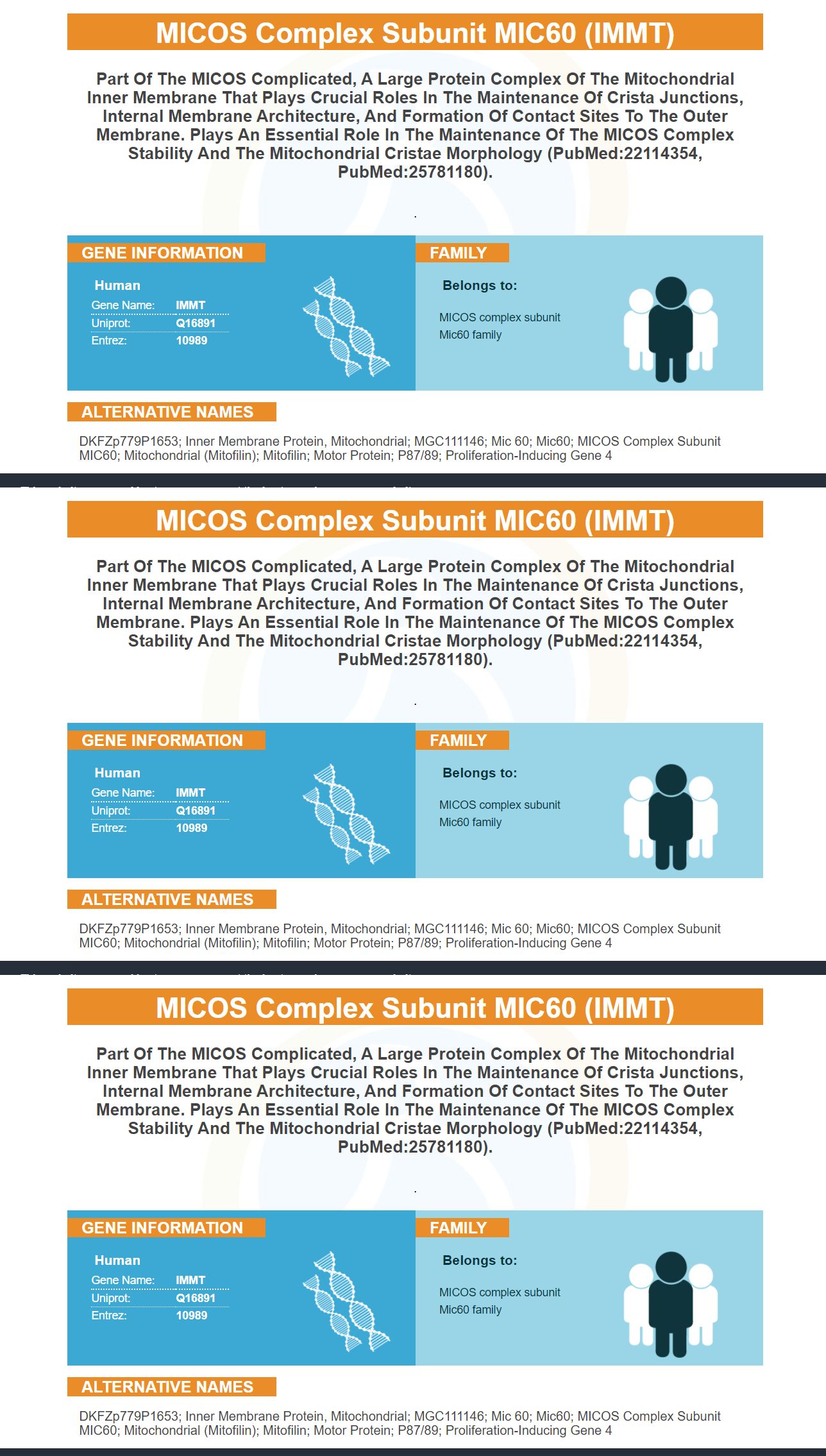
Facts about MICOS complex subunit MIC60.
.
Human | |
---|---|
Gene Name: | IMMT |
Uniprot: | Q16891 |
Entrez: | 10989 |
Belongs to: |
---|
MICOS complex subunit Mic60 family |
DKFZp779P1653; inner membrane protein, mitochondrial; MGC111146; Mic 60; Mic60; MICOS Complex Subunit MIC60; mitochondrial (mitofilin); mitofilin; motor protein; p87/89; proliferation-inducing gene 4
Mass (kDA):
83.678 kDA
Human | |
---|---|
Location: | 2p11.2|2 |
Sequence: | 2; NC_000002.12 (86143932..86195770, complement) |
Mitochondrion inner membrane; Single-pass membrane protein. Mitochondrion.
The MICOS complex includes the IMMT marker. The MICOS complex is a group of protein components inside the mitochondrial inner membrane. This article will provide information about MIC60, its uses, and other pertinent information. It is a useful marker for detecting IMMT activation. It can be bought from Boster Bio (or a trusted supplier). Regardless of which type of IMMT marker you need, Boster Bio can help you.
IMMT is a protein found in mitochondria that is a member of the MICOS complex. Its function is to regulate mitochondrial architecture. It is nuclear encoded and plays an important role in the formation of crista junctions. Boster Bio has developed an anti-IMMT antibody that reacts with samples of different species. Boster Bio antibodies react with IMMT samples from many animals. The company has developed antibodies to the proteins using mouse and rabbit models. Subunits MIC60 of the MICOS Complex play an important role in the morphology of mitochondrial cristae and inner membrane.
MIC60 is a multiprotein protein complex that is essential in the structure and functioning of mitochondria. It has been linked to several neurodegenerative diseases and may be a key player in the health of post-mitotic mitochondria-dependent neurons. Mic60 deficiency is associated with several neurodegenerative conditions, including fetal Down syndrome.
MIC60 is a key component of the mitochondrial inner membrane. It is also a component in the MICOS (Microorganic inner membrane protein) and MIB (Microorganic intracellular protein complexes), which bridge inner-membrane cristae cristae cristae cristae junctions. MIC60 also regulates ATP synthesise F0-F1 (cristae morphology) Despite its important role in mitochondrial metabolism, it is not required for several diseases and organ systems.
The MIC60/mitofilin Complex is a multiprotein compound of the mitochondrial inner cell that is responsible the structure of mitochondria. Mic60 can be found in more than half of all cells. Its distribution bands appear twisted and resemble a helix. Mic60 is expected spread into ring structures that have gaps. However, a twisted Mic60 signal is not visible in all mitochondria.
Mic60 is a unique pattern found in yeast cells. This makes it difficult to quantify individual groups. Mic60 immunolabeling yeast cell's mitochondrial tubules is complex and requires manual selection of imaged cells. Mic60 distribution is predicted to be affected significantly in yeast cells that lack the crista junctions.
Many copies of ATP synthase are found in the inner mitochondrial membrane, also known as F0F1ATPase. These proteins form a channel for protons, and they span the mitochondrial inner cell membrane. The F1 (or ATP synthase) component is made up five proteins and has a mollecular weight of about 360,000. The F0 is a multiprotein component that contains 8-15 small, c-subunits. The F1 complex combines movement of H+ with the synthesis of ATP.
Despite these effects, it has been shown that loss of Mic60 is associated with functional and structural disruption of neuromuscular junction synapses, as well as adverse effects on the health of axons and their mitochondria. These findings suggest that Mic60 may provide protection against multiple neurological disorders. In the meantime, Mic60's role in PD research remains unclear.
Mic60 can be found in different cell types and in opposing bands. Cristae in silico do not span the matrix space but instead form opposing distribution channels. Mic60 clusters are often formed in opposing pairs. Mic60 clusters in opposing pairs in the inner membrane of mitochondrial cells. Its morphology will also determine whether Mic60 clusters develop in the inner mitochondrial cells.
MIC60, a component of MIC60, plays a crucial role in mitochondrial structure and respiration. Loss of Mic60 can lead to a decline in cell viability. Mic60 overexpression is protective in an experimental toxicant model of PD. Exogenous DA is protected by Mic60 overexpression, but it is not clear why.
MIC60 can form clusters in different types human cells. It can also adopt a helical configuration. The mic60 distribution bands are shifted by 500 nm, and the Pearson correlation coefficient is almost zero, indicating that the two components are not correlated. Mic60 forms a distinct MICOS organizing center, in which it interacts other proteins. Mic60 can bind to different MICOS proteins, including the MIC10 and Mic12, ensuring that the complex forms two independent MICOS organizing centers.
Mic60 can lead to changes in mitochondrial function due to its vulnerability to oxidative damage. This happens because the mitochondrial environment is high in ROS. Photodynamic therapy decreased the Mic60 protein level in cultured cells. Homoharringtonine also increases Mic60's mRNA expression, which is unusual for this gene. These results suggest that Mic60 is an integral component of the MICOS complex.
Mic60 is not required to locate at the cristae junctions in mice. Mic60 shares similar properties to MICOS cells in terms morphology and mtDNA. Mic60 is located at the cristae junctions. It promotes the formation of Mic27. Mic27 promotes the mic60-mic27 assembly, which also helps the mitochondrial structure.
Numerous MIC60 components can be found in microtubules. These molecules are involved in regulating the structure and function of mitochondria. Mic60 is an attractive therapeutic target in mitochondrial disorder. Mic60 is an exciting therapeutic target in mitochondrial dysfunction. Its deficiency has been shown to be linked to cellular stress resulting from PD. Mic60 rescues PD phenotypes of PINK1 mutant Drosophila. Mic60 deficiency might be an effective therapeutic target in multiple neurological disorders.
Mic60 is necessary for stability of Mic19. Mic27 is found in more prominent mitochondrial foci. Mic60 is also found within the mitochondria. This may indicate that there is a second MICOS organization center. While the distribution of Mic60 is not uniform, it is distributed within mitochondria. It may have an intrinsic ability to self-assemble, as demonstrated in the MICOS studies.
Although the molecular basis of Mic60 assembly is not known, it is essential for proper organization and maintenance of the inner membrane. The outer membrane associated determinants may direct the biogenesis and assembly of Mic60. Mic60 interacts with the mitochondrial sorting and import machinery complex, which may facilitate proper spacing of the inner boundary and proper organization of the outer membrane components. The MIC60-MIC60 complex may also be involved in mitochondrial sorting machinery and import.
PMID: 8039717 by Icho T., et al. A novel human gene that is preferentially transcribed in heart muscle.
PMID: 9168817 by Gieffers C., et al. Mitofilin is a transmembrane protein of the inner mitochondrial membrane expressed as two isoforms.