This website uses cookies to ensure you get the best experience on our website.
- Table of Contents
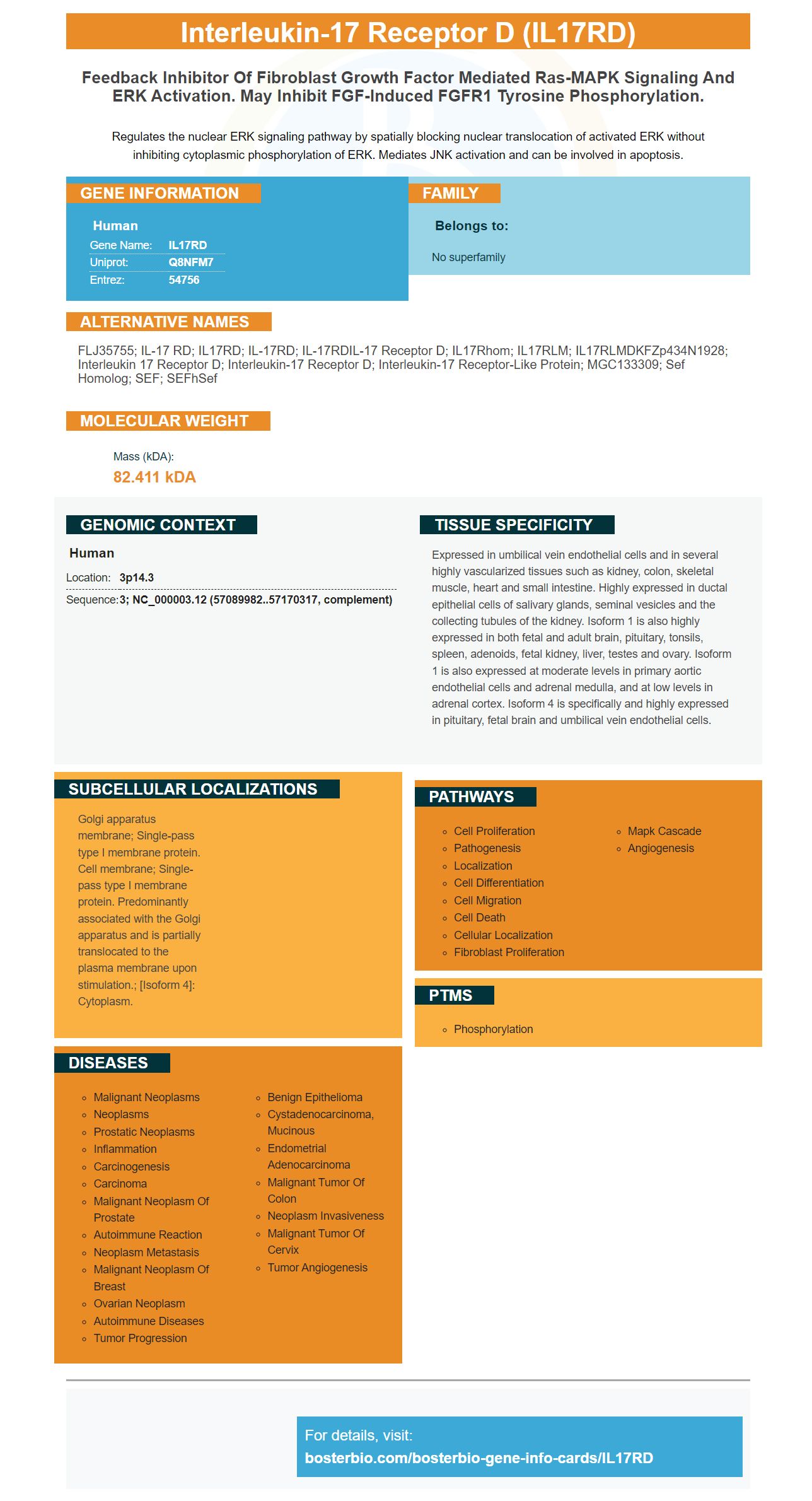

Facts about Interleukin-17 receptor D.

Regulates the nuclear ERK signaling pathway by spatially blocking nuclear translocation of activated ERK without inhibiting cytoplasmic phosphorylation of ERK. Mediates JNK activation and can be involved in apoptosis.
| Human | |
|---|---|
| Gene Name: | IL17RD |
| Uniprot: | Q8NFM7 |
| Entrez: | 54756 |

| Belongs to: |
|---|
| No superfamily |

FLJ35755; IL-17 RD; IL17RD; IL-17RD; IL-17RDIL-17 receptor D; IL17Rhom; IL17RLM; IL17RLMDKFZp434N1928; interleukin 17 receptor D; interleukin-17 receptor D; Interleukin-17 receptor-like protein; MGC133309; Sef homolog; SEF; SEFhSef
Mass (kDA):
82.411 kDA

| Human | |
|---|---|
| Location: | 3p14.3 |
| Sequence: | 3; NC_000003.12 (57089982..57170317, complement) |
Expressed in umbilical vein endothelial cells and in several highly vascularized tissues such as kidney, colon, skeletal muscle, heart and small intestine. Highly expressed in ductal epithelial cells of salivary glands, seminal vesicles and the collecting tubules of the kidney. Isoform 1 is also highly expressed in both fetal and adult brain, pituitary, tonsils, spleen, adenoids, fetal kidney, liver, testes and ovary. Isoform 1 is also expressed at moderate levels in primary aortic endothelial cells and adrenal medulla, and at low levels in adrenal cortex. Isoform 4 is specifically and highly expressed in pituitary, fetal brain and umbilical vein endothelial cells.
Golgi apparatus membrane; Single-pass type I membrane protein. Cell membrane; Single-pass type I membrane protein. Predominantly associated with the Golgi apparatus and is partially translocated to the plasma membrane upon stimulation.; [Isoform 4]: Cytoplasm.


If you're looking for a quality primary antibody for IL17RD, you've come to the right place. Boster produces high affinity antibodies that are frequently cited in the scientific community. Boster antibodies have been validated and characterized for Western Blotting, Immunohistochemistry, and ELISA. With Boster, you can rest assured that your research will be as productive as possible.
Boster Bio Anti-Interleukin 17Receptor D IL17RDMarker is a monoclonal anti-human IL17 antibody. This antibody has been tested for WB. It can be stored at -20C or 4degC up to one month. It reacts with Rat, Mouse, Human. Refer to the product label for further information.
The membrane-bound long version of human IL-17 RD is composed of a 27aa signal peptide and a 272aa intracellular domain. It also contains a 20aa transmembrane section. This product exhibits a TRAF6 binding motif. Before use, the dilution should be optimized. Keep the product at 4°C until you use it.
This antibody is used in the treatment of autoimmune diseases. It is the only antibody to target the second IL-17 receptacle. Although many other IL-17 receptors are known, the discovery of a new one is both exciting from a clinical and basic perspective. This new drug has not been approved for clinical use. The research will continue to support the use of the anti-IL-17R D IL17RD Marker in psoriasis research.
To determine the binding affinity between human IL17C and mouse IL17RE/Fc, IL17RE/Fc interaction tests were conducted. To identify antibodies that inhibit the binding of IL17C to IL17RE/Fc, chimeric human and mouse IgG2a antibodies were used. The binding was detected by Streptavidin-ECL.
IL-17/IL-17R mediates cellular activity through PI3K/MAPK signaling pathways. Recent research investigated the role played by the JAK2/STAT3 signaling pathway during IL-17-mediated self-destructive autophagy. Oxaliplatin-treated cells in SMMC-7721 cells had higher levels than control cells of p–JAK2 protein and p–STAT3 protein.
High-affinity primary antibodies against IL17RD make a great choice for many immunoassay applications. These antibodies are composed two identical arms. The Fv is a variable area and the CL is a constant one. Both of these arms are responsible for the direct effect of the antibody's binding to and recognition of the target protein. This class of antibodies may have different effector mechanism.
The monoclonal antibody used in research was developed in mouse models of autoimmune disorders and Rheumatoid arthritis. They have been validated using multiple cell types in clinical trials. They have been proven effective in multiple clinical trials using mouse models of rheumatoid, and Th27-mediated murine models of Multiple Sclerosis. They are also effective in treating sepsis.
Antigen-specific antibodies are produced by immune cells when they encounter foreign proteins. These antibodies are key components of a successful immunotherapy. High-affinity memory B cells develop in special lymphoid structures called germinal centers. These germinal center are found in lymphoid tissues including bronchus. They are carriers of the germinal center marker, GL7, as well as a variety other markers. Autoantibodies with high affinity are produced by high-affinity memory cells B cells.
The IL17R receptor is a protein that has two main types: IL-17A/F (or IL-17RD). Both proteins are full-length polypeptide chains of amino and nucleic substances. FIGS. FIGS. The nucleotide symposia are displayed in bold and underlined fonts.
The activation of the immune systems is promoted by cytokines IL-17 and their receptors. Various antibodies are available to target these cytokines. Both from a clinical and a basic perspective, the discovery of IL-17RD is very exciting. This discovery could be a therapeutic intervention in the context of autoimmune diseases. The research is still in its infancy. It is already helping to understand the role played by IL-17 in autoimmune disorders.
An innovative use of the IL17RD mark is monitoring MS patients who are receiving an IL-17 antagonist. The blood is tested for IL-17 RNA transcripts, as well as protein products to determine their expression levels. If the gene expression is elevated relative to a control sample, the patient should continue the treatment with IL-17 antagonists. This method can be used to diagnose MS patients or to administer IL-17 antagonist therapy.
Antibodies that recognize IL-17 can bind to IL-17 receptors to allow them to recognize cancer cells. The IL-17RD marker can also be used to profile gene expression. It can be used either alone or in combination with other methods for identifying tumor resistance markers. How can the IL-17RD marker help in clinical practice? There are several ways to test the IL-17 receptor, but one method is known to be the most effective and versatile.
A series functional experiments validate the IL17RD markers availability. The cytokine IL17A has high specificity, and affinity. It is produced by activated T cells and plays a pivotal role in the pathogenesis of multiple autoimmune diseases. The Boster antibody binds this cytokine, validating its specificity.
Availability of the IL17RD marker is available as part of a large catalog of biological assays. This antibody can be used in a variety of animal samples. It is polyclonal, and reacts with Interleukin-17 R receptor D. Boster Bio offers a wide range of antibodies against the IL17RD mark. Boster Bio's IL17RD antibodies are derived from rabbits and mice. The IL17RD is a key component in the immune system. It mediates the activation JNK. It is also involved apoptosis.
Diverse innate immune cell types produce IL-17. This includes NKp46+NK Cells, intestinal Paneth and neutrophils. It secretes IL-17A, and IL-17F. Human entheses also contain CD4+ and CD8+ cells that produce IL-17A as well as TNF-a. IL-17RD plays an important role in the detection SpA and other autoimmune disorder.
In the treatment of autoimmune disorders, antibodies that target IL-17 cytokines and IL-17 receptors can be used. Boster bio now has the IL17RD marker. This is a great development. This marker can be used for clinical trials. It is essential that the protein is present in order to diagnose the disease. Antibodies against IL-17R can be used to help doctors diagnose underlying causes of psoriasis.
The IL17RD cytokine is a cytokine which associates with IL17RA/IL17RC and modulates IL17A signaling context-dependently. It can interact with other IL17 receptors, and affect signaling pathways downstream from other IL17 cytokines. This marker is currently only available for human use. However, it is expected that its price will drop over time.
The IL17RD molecule binds a region of the NFKB pathway, the Cterminal nuclear localization sequencing. This interaction is located at residues 321-395. It may be used to inhibit activation the NFKB pathway. It is also an scaffolding protein that binds directly to the SEFIRdomain, where it forms a p50 binding domain.
Besides its regulatory role in the inflammatory response, IL17RD also regulates crosstalk between osteoclasts and synovial cells, two important cell types in arthritic disease. It is also believed to regulate bone remodeling during arthritis. It has also been implicated by several mouse and human studies. It is still unclear how it affects these types of cells. Multiple pathways are connected to the IL17RD marker including the ERK pathway.
PMID: 12958313 by Xiong S.Q., et al. hSef inhibits PC-12 cell differentiation by interfering with Ras- mitogen-activated protein kinase MAPK signaling.
PMID: 14742870 by Preger E., et al. Alternative splicing generates an isoform of the human Sef gene with altered subcellular localization and specificity.


