This website uses cookies to ensure you get the best experience on our website.
- Table of Contents
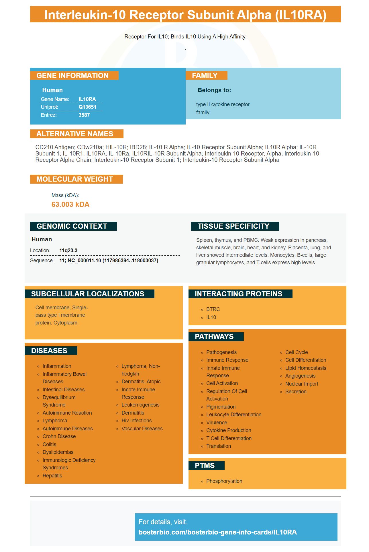
Facts about Interleukin-10 receptor subunit alpha.
Receptor for IL10; binds IL10 Using a high affinity.
.Human | |
---|---|
Gene Name: | IL10RA |
Uniprot: | Q13651 |
Entrez: | 3587 |
Belongs to: |
---|
type II cytokine receptor family |
CD210 antigen; CDw210a; HIL-10R; IBD28; IL-10 R alpha; IL-10 receptor subunit alpha; IL10R alpha; IL-10R subunit 1; IL-10R1; IL10RA; IL-10Ra; IL10RIL-10R subunit alpha; interleukin 10 receptor, alpha; interleukin-10 receptor alpha chain; Interleukin-10 receptor subunit 1; interleukin-10 receptor subunit alpha
Mass (kDA):
63.003 kDA
Human | |
---|---|
Location: | 11q23.3 |
Sequence: | 11; NC_000011.10 (117986394..118003037) |
Spleen, thymus, and PBMC. Weak expression in pancreas, skeletal muscle, brain, heart, and kidney. Placenta, lung, and liver showed intermediate levels. Monocytes, B-cells, large granular lymphocytes, and T-cells express high levels.
Cell membrane; Single-pass type I membrane protein. Cytoplasm.
IL-10 is an important part of our immune system. IL-10 has many beneficial functions. It can suppress inflammation and increase cytokine release. However, its most important use is the regulation of immune responses in the body. This article will be devoted to the role played by IL-10. It will also give information about how this cytokine can be produced by cells. This could lead to its use in antigens.
Boster Bio's IL10 is produced using a His-tagged, pCEPV19 vector. This recombinant cytokine has been detected using ELISA. It inhibits inflammation by blocking pro-inflammatory cytokines. Reactive microgliosis can occur when IL-10 production is not controlled.
The biological mechanisms responsible for regulating cytokine expression are similar in aging, differentiation, and cell-death. It shares some characteristics with the innate immune response and the inflammatory response. Therefore, Boster Bio IL10 may have therapeutic potential in the treatment of inflammatory disorders. Further research is needed to identify whether Boster Bio IL10 promotes the phosphorylation of STAT3 in THP-1 cells and in macrophages.
IL-10 has a very short half-life in vivo, and the stability of the non-covalent dimer is dependent on pH and temperature. It dissociates into a monomer at low protein concentrations and acidic pH. Acidic pH is also a problem for hematomas and skin irritations caused by fractures. Boster Bio created a supplement that increases the production of IL-10.
Boster Bio IL10 is a pleiotropic cytokine with anti-inflammatory properties. It interacts with the IL-10 receptor, which is expressed most frequently on monocytes. By activating the IL-10 receptor, IL-10 inhibits the production of pro-inflammatory cytokines. IL-10 has several benefits for promoting healthy aging, and it has a very important role in fighting infections and other diseases.
This product is made from a synthetic polymer, which links the C-terminus and N-terminus of each IL-10 monomer. The polymer is stable and a molecule made by this method has a higher bioactivity than natural mouse IL-10. This product also has the potential to increase IL-10 production. It has also been shown to improve the immune system, as it is able to fight bacterial infections.
This product does, however, not inhibit IL-10R1 receptors. You can also inject anti-IL-10R1 receptor antibodies to block IL-10Dimer formation. It is believed that dimer is involved with the interaction between IL-10R (IL-10R) and IL-10R. For dimerization to activate IL-10R, the receptor binding domain of IL-10 must be present.
The IL-10 genes is involved in the creation of a multifunctional cytokine. It also regulates cell adhesion, acute phase protein and glucose homeostasis. Polymorphic genotypes within the IL-10 gene have been associated with an increased incidence of chronic diseases like T2DM and autoimmune conditions. The results of the study also indicate that the Boster Bio IL10 is effective in the treatment of diabetic patients.
In addition to its anti-inflammatory properties, IL-10 is also involved in controlling viral immunity. Studies on LCMV show that it inhibits proinflammatory responses in both the innate and adaptive immune mechanisms. Furthermore, IL-10 inhibits tissue damage due to an exacerbated adaptive immune response. As a result, IL-10 is a common target for viral infections. This immunoregulatory property is used to treat infections that persistently recur.
In the current study, we investigated whether LPS-treatment of RAW/NC cells increases production of Cytokines. RAW264.7 cells, seeded at a density 1.55 x 10⁵ cells/mL in 24-well plates were used. Cells were pretreated for 1 h with PCA1 (80 mgmM) and incubated for 6 h with LPS (1mg/ml). Fluorescence images were acquired using a confocal microscopy system.
LPS stimulates macrophages, and increases intracellular calcium. Calcium is also involved transcriptional activation in inflammatory cytokines, chemokines. In this study PCA1 significantly inhibited LPS inducing ROS generation by RAW264.7 cells. PCA1 also decreased intracellular ROS levels. PCA1 promoted degradation of IKKa/b, which in turn reduced the production and release of cytokines.
LPS treatment of RAW/NC-cells also increased IL-6 and IL-10 production. Interestingly, Stat1 and Ahr inhibit the signaling of LPS in RAW/NC cells and macrophage-like cells. These results support Ahr's inhibition in the production proinflammatory cytokines. LPS induces IL-6 (and Ahr) in RAW/NC cellular cells.
LPS stimulates both NFkB activation and YAP. Both proteins play a role in the innate immunity response. Lactate also suppresses NFkB activation by macrophages. Further research will be done into the effects that lactate can have on LPS-stimulated, NFkB. Our results suggest that LPS can induce cytokine production through two mechanisms: activation of GPCR81 and the nuclear translocation of NF-kB.
Multiple inflammatory mediators were produced in LPS-induced RAW/NC cell lines. TNFa, tumor necrosis Factor-a, and interleukin-6 are the major mediators of the immune reaction in these cells. These findings support the idea that sepsis can be caused by the presence these mediators. These mediators are crucial in the regulation of innate immune reactions.
The study also revealed that PCA1 had an impact on Keap1/Nrf2-signaling pathway. LPS treatment increased expression of HO-1, HMGB1 and HMGB1 proteins within RAW/NC-cells. PCA1 is also known to inhibit the translocation IkBa/b and increase cytokine release. The findings also revealed that LPS induced RAW/NC cell growth increased IL-1 as well as IL-6.
LPS treatment resulted in macrophage activation. Cell-conditioned media levels of NO and Nitrite were measured. A Griess assay was used to add 50 mLs of cell-conditioned medium to each well containing wortmannin, LPS, and other substances. The respective compounds were then added to the cells and cultured to determine the levels of nitrites or inflammatory cytokines.
LPS-stimulated macrophages also induce NFKB activation. This activation begins with binding of the NFKB subunit to p50. The complex then recruits to the IL-6 enhancer. This activity is inhibited by Ahr, but it does not affect LPS-stimulated levels of NF-kB-stimulated p65.
IL-10, a pleiotropic cytokine, has powerful anti-inflammatory and immunosuppressive capabilities. It inhibits production of MHC Class II molecules, reduces proinflammatory cytokines, and limits T cell proliferation. These properties are essential for maintaining good health and fighting infections. The role of IL-10 in the body is not yet fully understood, but this compound has potential to become a new treatment for inflammatory diseases.
Studies have shown that IL-10 inhibits inflammatory cytokines' production by decreasing immune system expression of RAAS. It inhibits tubular injury after UUO. It also decreases inflammatory and cytokine messenger RNA by inhibiting HuR. Moreover, exogenous IL-10 administration reduces tubular fibrosis. Exogenous IL-10 administration reduces tubulointerstitial fibrosis. This is due to upregulation of HMWHA.
Study of IL-10's role in atherosclerosis has been done. Atherosclerosis is a chronic inflammatory response that involves dysregulation of the ECM. This inflammation begins with endothelial disruption and recruitment of T lymphocytes to the site of injury. The arterial wall is then damaged by Macrophages, which are then transformed into lipid-laden cells called foam cells. In turn, these cells initiate the formation of atheromatous plaques.
IL-10 has also been implicated in obesity-related metabolic disorders. It also regulates the differentiation and proliferation of M1 macrophages. It also inhibits mTORC1 in macrophages. These results suggest that IL-10 plays an important role in sepsis associated encephalopathy. More research is needed to find out how IL-10 inhibits the inflammation. This article will explain the beneficial effects of IL-10 on the body.
The circulating IL-10 levels are associated with an increased sensitivity for immunosuppressive agents and antibiotics. High levels of IL-10 are also associated with immune suppression and disease progression. IL-10 administration can promote proliferation of cytotoxic T cells and increase IFNg production in rodent tumor models. This results in enhanced antitumor action. High levels of IL-10 in cancer patients can also induce systemic immune activation, as well as expansion of both tumor-infiltrating CD8+ T cells and systemic CD8+ cells.
IL-10 promotes adhesion to EPC and EPC migration. It also increases MMP-9, VEGF, and VEGF. It activates the STAT3 signaling pathway. Further studies of IL-10's effect on fibrosis may reveal new therapeutic targets. This is a great time to study this particular cytokine in the context regenerative medicine.
Inflammation is caused by the overproduction of cytokines from immune cells. This overproduction of cytokines can lead to tissue damage and organ failure. Understanding the role played by IL-10 in COVID-19 is key to better treatments. However, there are still many questions about how IL-10 functions and how it inhibits COVID-19 swelling.
PMID: 8120391 by Liu Y., et al. Expression cloning and characterization of a human IL-10 receptor.
PMID: 12133952 by Usacheva A., et al. Two distinct domains within the N-terminal region of Janus kinase 1 interact with cytokine receptors.