This website uses cookies to ensure you get the best experience on our website.
- Table of Contents
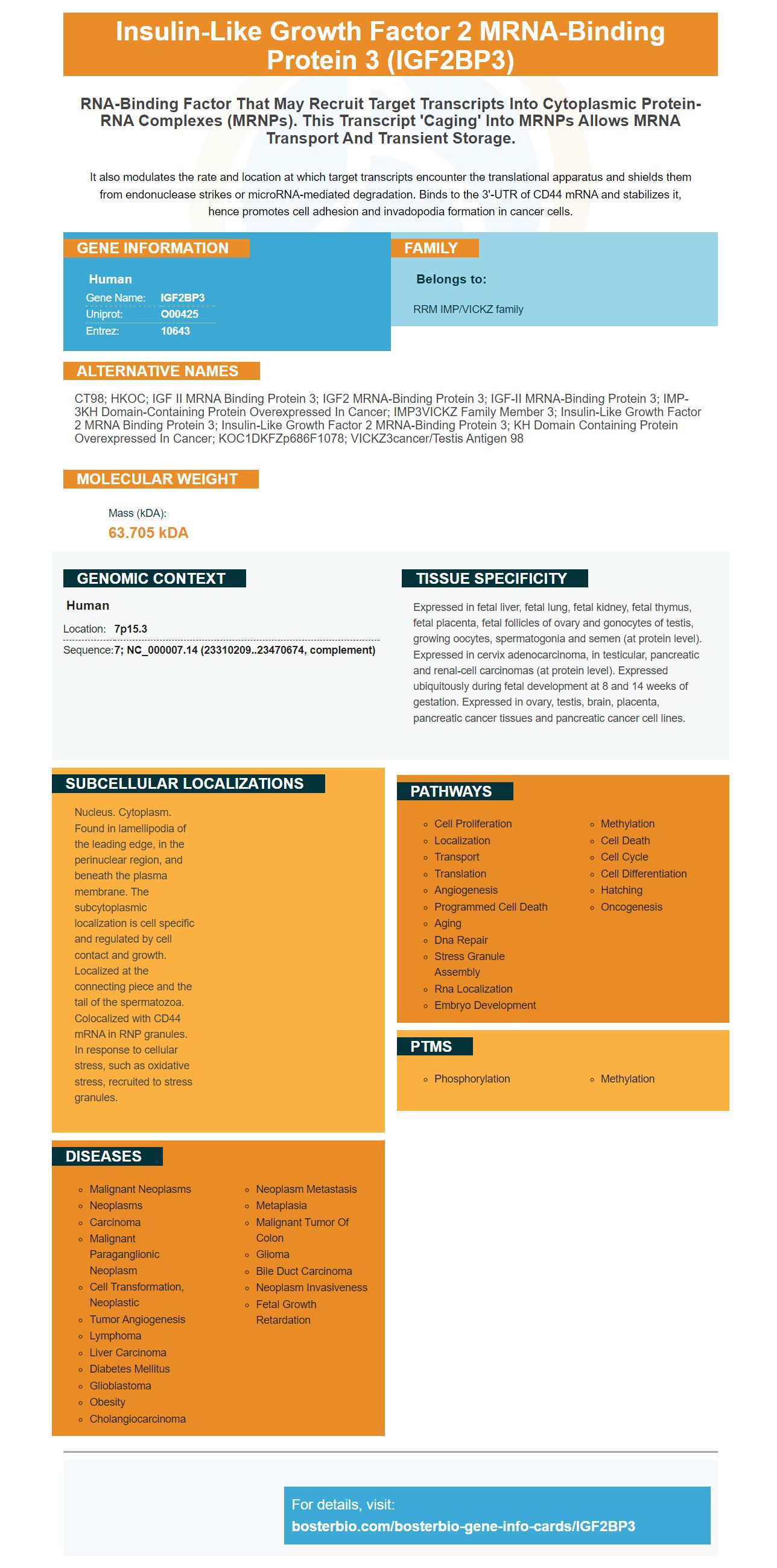
Facts about Insulin-like growth factor 2 mRNA-binding protein 3.
It also modulates the rate and location at which target transcripts encounter the translational apparatus and shields them from endonuclease strikes or microRNA-mediated degradation. Binds to the 3'-UTR of CD44 mRNA and stabilizes it, hence promotes cell adhesion and invadopodia formation in cancer cells.
Human | |
---|---|
Gene Name: | IGF2BP3 |
Uniprot: | O00425 |
Entrez: | 10643 |
Belongs to: |
---|
RRM IMP/VICKZ family |
CT98; hKOC; IGF II mRNA binding protein 3; IGF2 mRNA-binding protein 3; IGF-II mRNA-binding protein 3; IMP-3KH domain-containing protein overexpressed in cancer; IMP3VICKZ family member 3; insulin-like growth factor 2 mRNA binding protein 3; insulin-like growth factor 2 mRNA-binding protein 3; KH domain containing protein overexpressed in cancer; KOC1DKFZp686F1078; VICKZ3cancer/testis antigen 98
Mass (kDA):
63.705 kDA
Human | |
---|---|
Location: | 7p15.3 |
Sequence: | 7; NC_000007.14 (23310209..23470674, complement) |
Expressed in fetal liver, fetal lung, fetal kidney, fetal thymus, fetal placenta, fetal follicles of ovary and gonocytes of testis, growing oocytes, spermatogonia and semen (at protein level). Expressed in cervix adenocarcinoma, in testicular, pancreatic and renal-cell carcinomas (at protein level). Expressed ubiquitously during fetal development at 8 and 14 weeks of gestation. Expressed in ovary, testis, brain, placenta, pancreatic cancer tissues and pancreatic cancer cell lines.
Nucleus. Cytoplasm. Found in lamellipodia of the leading edge, in the perinuclear region, and beneath the plasma membrane. The subcytoplasmic localization is cell specific and regulated by cell contact and growth. Localized at the connecting piece and the tail of the spermatozoa. Colocalized with CD44 mRNA in RNP granules. In response to cellular stress, such as oxidative stress, recruited to stress granules.
The IGF2BP3 protein assembles in stress granules with target mRNAs to promote localization to regions of active translation. This protein is also known as nRNP, nuclear ribonucleoprotein, SG, and RISC, RNA-induced silencing complex. Here are some of the best uses of this protein.
Developed by BioRad, Cell Counting Kit-8 (CCK8) is a colorimetric assay kit for live cell counting that measures cytotoxicity and proliferation. Unlike other cell-based assays, it does not require lysing cells to obtain accurate results. Furthermore, its non-radioactivity means that it is easier to use and maintain than the standard radioactive incorporation assay.
The Kit is a ready-to-use solution that is stable at 4oC for 1 year. It can be completed at any time. While this kit is a convenient and reliable way to test cell viability in cancer patients, it cannot verify cell death. To determine cell death, Cytotoxicity LDH Assay Kit-12 (CK12) is recommended. The data obtained from this assay was provided by Reaction Biology Corporation. The measurement requires a microplate reader with a 450-490 nm filter.
Cell Counting Kit-8 reduces WSTS* to an orange-colored product that is non-radioactive and convenient to use. It reduces WSTS* by cellular dehydrogenases to produce formazan dye. The amount of orange formazan produced is directly proportional to the number of living cells. The amount of orange formazan produced is proportional to the number of living cells in the test sample.
Lung cancer is the most common type of cancer and causes the highest number of deaths worldwide. The 5-year survival rate is only 18% to 50%. Most lung cancers are non-small cell lung cancer (NSCLC). Despite the prevalence of advanced-stage NSCLC, the primary treatment remains surgical resection combined with chemotherapy or radiotherapy. Although this form of lung cancer has a low survival rate, treatment should be effective to decrease the likelihood of metastasis or recurrence.
The CCK-8 is a ready-to-use cytometric assay that is used to quantify the number of cells in a culture medium. It should be thoroughly mixed and stored in sterile conditions to prevent contamination. After mixing, the cell suspension should be incubated for at least 4 hours. To measure cell viability, the solution should be incubated for 0.5 to 4 hours at 37degC. Over-incubation can result in false results.
This assay requires a cell sample to be incubated at 0 to 5 degrees Celsius in a dark environment. The sample should be harvested at least 48 hours after incubation, and samples with few cells can be incubated up to 24 hours. The samples are then analyzed by measuring their absorbance at 450 nm with a microplate reader. OD values should be 0.8 to 1.5.
Cell Counting Kit-8 is an easy-to-use one-bottle solution for the quantification of live cells. It is a non-radioactive and colorimetric assay, allowing for quick, accurate, and convenient cell counting. It is also more affordable than other conventional cell counting methods. Using CCK-8 is easy and cost-effective, and your patients will thank you.
The Cell Counting Kit-8 is convenient and highly sensitive for performing cell viability assays. It is non-radioactive and uses a water-soluble tetrazolium salt (WST-8), which produces an orange formazan dye upon bio-reduction in the presence of an electron carrier. Using this kit eliminates the need for pre-mixing of components, reducing the amount of WST-8 to a yellow-orange solution. The resulting orange formazan product has an absorbance at 460 nm, which is directly proportional to the number of living cells.
CCK-8 has the ability to reduce WST(r)-8 in prostate cancer cells. The CCK-8 is compatible with 100 or 200-ml multichannel pipettes and can be stored for up to 6 months at room temperature. The kit is not stable in freezers and should be mixed thoroughly before incubation. It should never be stored for prolonged periods of time or frozen for long periods. The solution should be thoroughly mixed before use.
This cell counter is a convenient way to monitor the progression of breast cancer. It is available in various sizes and should be stored at a temperature of 0-5 oC and kept away from light. Before use, mix the kit thoroughly. Add the cells to the inner wells. The cell counter will then measure the cellular viability. If there are fewer cells in the sample, you can incubate the samples for 24 hours.
The cell counter uses the Cell Counting Kit-8 (CCK-8) to measure the number of cells in culture medium. Add 100 ul of culture medium to the Cell Counting Kit-8 and mix it well. Tap the plate to avoid bubbles. Then incubate the mixture for 0.5 to 4 hours at 37degC. Do not over incubate the cell counter because the resulting color may be incorrect.
The Selleck Cell Counting Kit-8 (CCK-8) is a non-toxic, water-soluble reagent that allows for sensitive colorimetric assays of cell viability. Its formula uses Dojindo's highly water-soluble tetrazolium salt, WST-8, which is reduced by cell dehydrogenase activities to yield a yellow-colored formazan dye. The solution is compatible with 10 ml and 200 ml multi-channel pipettes.
PMID: 9178771 by Mueller-Pillasch F., et al. Cloning of a gene highly overexpressed in cancer coding for a novel KH-domain containing protein.
PMID: 10525192 by Mueller-Pillasch F., et al. Expression of the highly conserved RNA binding protein KOC in embryogenesis.