This website uses cookies to ensure you get the best experience on our website.
- Table of Contents
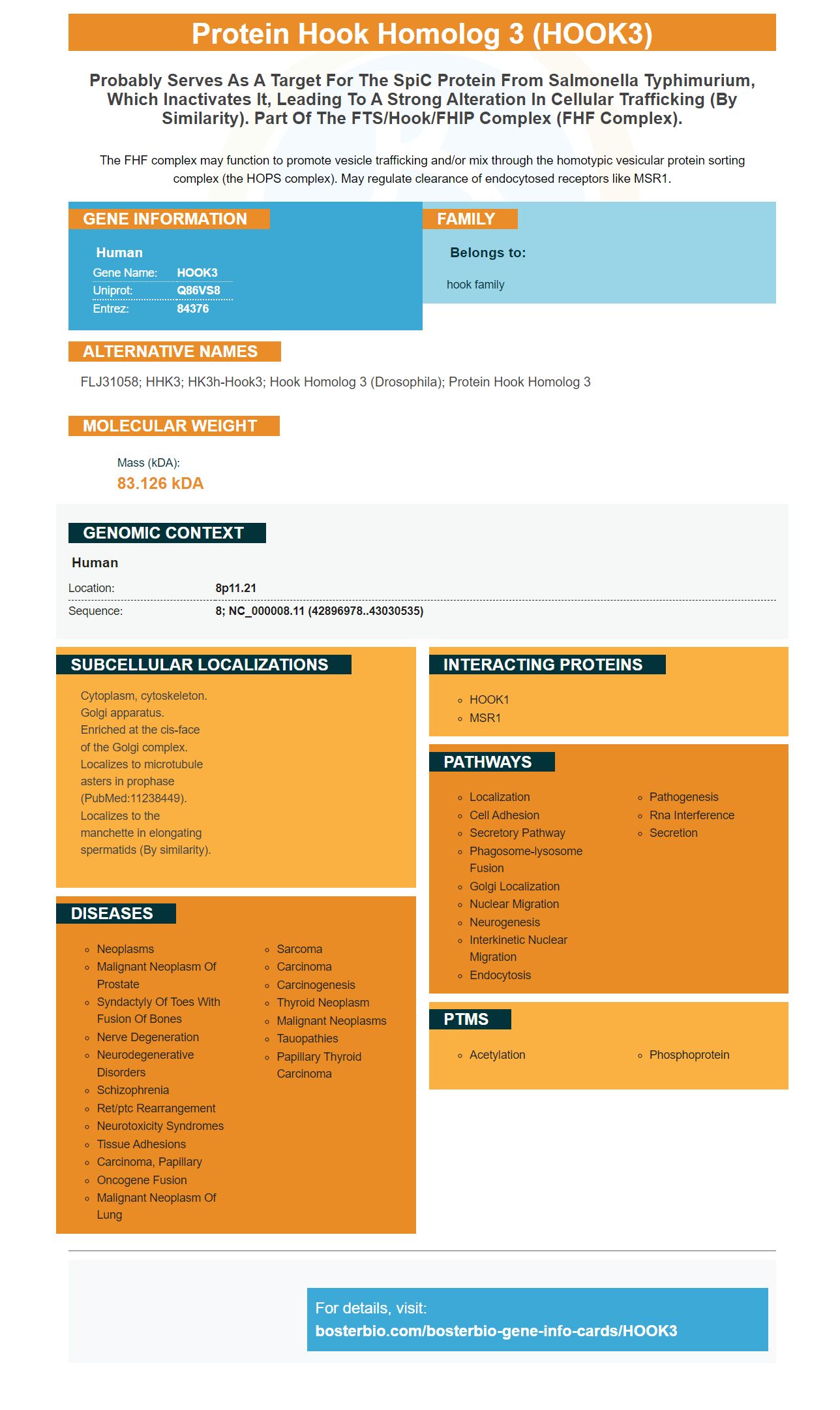
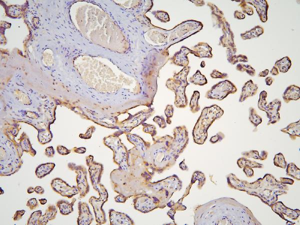
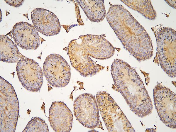
Facts about Protein Hook homolog 3.
The FHF complex may function to promote vesicle trafficking and/or mix through the homotypic vesicular protein sorting complex (the HOPS complex). May regulate clearance of endocytosed receptors like MSR1.
Human | |
---|---|
Gene Name: | HOOK3 |
Uniprot: | Q86VS8 |
Entrez: | 84376 |
Belongs to: |
---|
hook family |
FLJ31058; hHK3; HK3h-hook3; hook homolog 3 (Drosophila); protein Hook homolog 3
Mass (kDA):
83.126 kDA
Human | |
---|---|
Location: | 8p11.21 |
Sequence: | 8; NC_000008.11 (42896978..43030535) |
Cytoplasm, cytoskeleton. Golgi apparatus. Enriched at the cis-face of the Golgi complex. Localizes to microtubule asters in prophase (PubMed:11238449). Localizes to the manchette in elongating spermatids (By similarity).
What is HOOK3 exactly? A cytosolic, coiled-coil proteins. It performs a variety of functions in cells, including the recognition of cell membrane proteins and the processing of food ingredients. Its cytosolic location, which is essential for many reasons including its ability temperature and pH to sense, is vital. Scientists who study HOOK3 can also use the marker in other applications, such as cancer research.
The Protein Hook homolog 3, encoded by the HOOK3 protein gene These proteins possess conserved N terminal domains, as well as divergent Ctermini. The Drosophila hook protein is a component of the endocytic compartment. Boster Bio's HOOK3 antibody recognizes the amino acids 210 to 260 of human HOOK3.
The HOOK3 protein expression assay is highly specific and has many applications. Boster's antibodies have been highly cited and validated on Western Blotting and Immunohistochemistry. They are a perfect choice for a variety of applications. Researchers can use HOOK3 as a tool to detect tumor cells and cancer metastases and monitor inflammation.
HOOK3 can be found in many different tissues in mammals. Hook3 is also expressed in the brain and skeletal muscles. Hook3 can also appear in the liver as well as the kidney and HEK293-cells. Molecular structures of HOOK3 indicate that it is found in all organs including the brain and liver.
HOOK3 (cytoplasmic coiled coil protein) interacts with LIC1 to form a hydrophobic cleft. It is possible the Hook domain may interact with LIC1's second helix. The binding interface of the Hook domain is not well conserved. Therefore, the mutant MBPLIC1F447A failed LIC1 binding.
A variety of resources can be used to identify microRNAs that target HOOK3. Roadmap Epigenomics database can help you identify transcription factors that regulate HOOK3 expression in different cell types. To study the regulation of HOOK3, you can also use the HOOK3 genes in the context microRNAs. Roadmap Epigenomics contains many databases that detail gene expression profiles and other functional features of HOOK3-protein.
Molecular analysis of HOOK3 revealed that it is associated to microtubules in cells and is expressed by different organelles. When HOOK3 was overexpressed, the HOOK3/LIC1 complex was found able to bind directly to microtubules. In vitro, HOOK3 has been shown to be associated in Golgi membranes. While in vivo, it is enriched in the cisGolgi.
The Hook domain is HOOK3's canonical seven-helix calponin homology (CH), fold that is similar to a holotype (Hook3). It also has an additional helix at it's C terminus. The Hook domain also has a hydrophobic cleft, which acts as a binding site to LIC1 helix-1. However, it is not yet fully characterized.
In a study using rotary shadowing electron microscopy, HOOK1 was found to be a kinesin-like protein with two well-separated globular domains on one end and a short neck-like region connecting them to a long, thin rod. These features are consistent with the N-terminal Hook domain, unstable CC1 region, and central Spindly motif12 motif.
Recent research revealed that the class A scavenger-receptor (SR-A), can interact with the cellular coiled coil protein HOOK3. However, the precise role of this coiled-coil protein in Golgi transport is still unknown. Hook3 might also be involved with the turnover of endocytosed Scavenger Rectors.
The C-terminal region of HOOK3 binds kinesin. The FHF complex promotes the distribution and distribution of AP-4 compounds to the perinuclear region. The NIN family is also associated with microtubule ends and is enriched in GO terms related kinesin motors.
The RET protooncogene and a HOOK3 coiled coil protein fusion created the HOOK3 genome. Researchers have discovered a new form RET/PTC rearrangement. This rearrangement was previously characterized in the expression gene products corresponding with the RET/PTC. It displayed gene expression signatures typical for RET/PTC, but was not positive for other RET rearrangements.
PMID: 11238449 by Walenta J.H., et al. The Golgi-associated hook3 protein is a member of a novel family of microtubule-binding proteins.
PMID: 17237231 by Sano H., et al. The microtubule-binding protein Hook3 interacts with a cytoplasmic domain of scavenger receptor A.