This website uses cookies to ensure you get the best experience on our website.
- Table of Contents
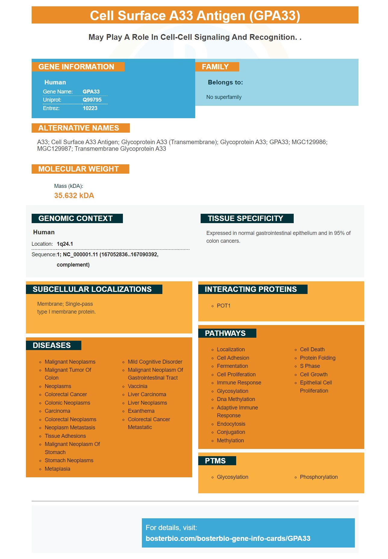
Facts about Cell surface A33 antigen.
Human | |
---|---|
Gene Name: | GPA33 |
Uniprot: | Q99795 |
Entrez: | 10223 |
Belongs to: |
---|
No superfamily |
A33; cell surface A33 antigen; glycoprotein A33 (transmembrane); Glycoprotein A33; GPA33; MGC129986; MGC129987; transmembrane glycoprotein A33
Mass (kDA):
35.632 kDA
Human | |
---|---|
Location: | 1q24.1 |
Sequence: | 1; NC_000001.11 (167052836..167090392, complement) |
Expressed in normal gastrointestinal epithelium and in 95% of colon cancers.
Membrane; Single-pass type I membrane protein.
A recent study described GPA33 expression in human peripheral leukocytes. This study could serve as a reference point for further research. Although it suggests that CD4+T-cells not committed in some way to a subset may be high expressors, the paper doesn't confirm this hypothesis. The study is a valuable resource paper. However, it needs to be more precise in its descriptions of function.
This investigation investigated the safety and efficacy for patients undergoing kidney transplantation. The cells were derived form human peripheral blood and cultured for 14 consecutive days in a laboratory using antiCD3/CD28 covered beads and IL-2. The results showed that patients' Tregs expanded in these experiments. Only two patients were unable or unable to expand their Tregs. There were three serious adverse events reported in the trial, including one death, but none were related to the cell infusion itself.
Tregs are derived mainly from peripheral blood, umbilical cord blood and thymus and have been recently isolated on a large-scale. To detect Tregs, peripheral blood samples have not been tested since the late 1990s. CliniMACS allows the isolation of cells from peripheral blood via a magnetic enrichment method. The system depletes CD8+ and CD19+ cells while enriching the CD25+ fraction. CliniMACS has been used by many groups to isolate Tregs.
While these cells do play a pivotal role in peripheral tolerance, they also limit inflammatory responses and induce tissue repair through amphiregulin production. Regulatory T cells have numerous reported functions, including protecting the body from allergic disorders, atherosclerosis, and tumor rejection. These cells may also control the immune system and regulate metabolic disorders. They are an important part of the immune system.
Tregs are involved in the immune system, suppressing different types cells. They produce granzymes that inhibit the proliferation of NK cells and secrete antiinflammatory chemicals. They also block IL-2's activity by sequestering the microenvironment. They also suppress the proliferation of NK cells by preventing their IL-g secretion. Moreover tTregs cells inhibit the production of IL-5 as well as IL-13.
T-bet's anti-inflammatory effect is closely connected to the tTreg-cell function. The antiTNF Alpha Monoclonal antibody is compatible and capable of recognizing many proteins ex vivo. They also prevent tumor-infiltrating immune cells from developing. It is important to consider whether your tTreg cells are functionally compatible with your own immune system.
In addition, the antigen-specific Treg cells in Boster Bio are more effective in suppressing alloimmune responses than their polyclonally expanded counterparts. These cells could also be useful in other ways. This treatment has an anti-CD40 receptor-mediated expansion ability. This makes it compatible with anti-CD3 Ab treatment.
The presence CD25 on tTregs cells increases the body's antitumor activity. Tregs are responsible for anti-tumor immunity, which decreases the cytotoxic ability of NK and NK cells. It is important to suppress immune responses in order to prevent the development and progression of autoimmune diseases. How can we identify human tTreg cell?
The major breakthrough in colon cancer research was the discovery of the gene that encodes the GPA33 protein in a cell from a colorectal cancer patient at Johns Hopkins University. GPA33 is highly expressed in colorectal carcinoma. A low level of PPARg kinase induces the gene. To confirm the results of this gene, the scientists used the irreversible PPARg antagonist (WY14643) and the Ciglitazone agonist (ciglitazone).
Rosiglitazone is a cis p –actin kinase kinase inhibitor. It inhibits PPARg induced KLF4 and a cellular transcription element that regulates GPA33. The present study involved treating colon cancer cells with rosiglitazone (10 mM) and DMSO. 24 hours later, RNA was extracted. The real-time PCR was used to detect FABP1, Cyclin D1, Keratin and Cyclin D1. The average of the results from three experiments was used.
GPA33, also known by cell surface A33 antigen (cell surface A33 gene), is expressed in the normal colonic epithelium and small bowel epithelium. Studies have shown that 95% of colon carcinomas are expressing the gene. It is expressed in colon cancers and has been shown to be strong in well-differentiated CRC tumours in IHC studies. Radioimmunotherapy is possible for GPA33-positive CRC tumours by using antibodies directed against the GPA33 genes.
These antibodies were made by combining FITC (131I) to label the antibody. Flow cytometry analysis revealed that A33scFv-Fc had high binding rates to GPA33-positive cells and low binding rates to GPA33-negative cells. This antibody was also shown specifically to bind the GPA33 protein, as demonstrated by its high binding rates with GPA33-positive tumor tissues. FITC–A33scFvFc also binds the GPA33 antibody, but does not bind to GPA33 negative liver tissue.
These results have been used in other studies that show the importance of GPA33 to colon cancer. Both the markers CDX2 (and GPA33) co-localize in intestinal epithelia and are known to be associated with T stage. They were also associated with the existence of LI-cadherin, which was found in tumor tissue. GPA33 expression was also associated with tumour-free resection margins, according to the findings.
This antibody targets GPA33 in colon cancers. It can visualize cancers that don't express GPA33. The antibody also accumulates in tumor grafts in LS174T tumor-bearing mice. It can also be used to guide the removal of colon tumor tissue. The antibody can be used in many other ways than GPA33.
Previous studies have shown that gpA33 has a high correlation with CDX2 or LI-cadherin in colon carcinoma. The positive control used normal colonic tissue as the control. Colon cancers are considered positive when more that 5% of the tumour cells are stained. The researchers have therefore concluded that GPA33 is an important marker of colon cancer. Further studies are needed to confirm the correlation and confirm if GPA33 is related to colon cancer.
Evidence suggests that PPARg is indirectly responsible for GPA33 regulation. Rosiglitazone activation of PPARg causes a small increase GPA33 expression. This effect is temporary and was eliminated when rosiglitazone was administered to cells. Interestingly, the same results were obtained in the absence of rosiglitazone, which is a PPARa receptor agonist.
PMID: 9012807 by Heath J.K., et al. The human A33 antigen is a transmembrane glycoprotein and a novel member of the immunoglobulin superfamily.
PMID: 9245713 by Ritter G., et al. Characterization of posttranslational modifications of human A33 antigen, a novel palmitoylated surface glycoprotein of human gastrointestinal epithelium.