This website uses cookies to ensure you get the best experience on our website.
- Table of Contents


Facts about Growth factor receptor-bound protein 7.

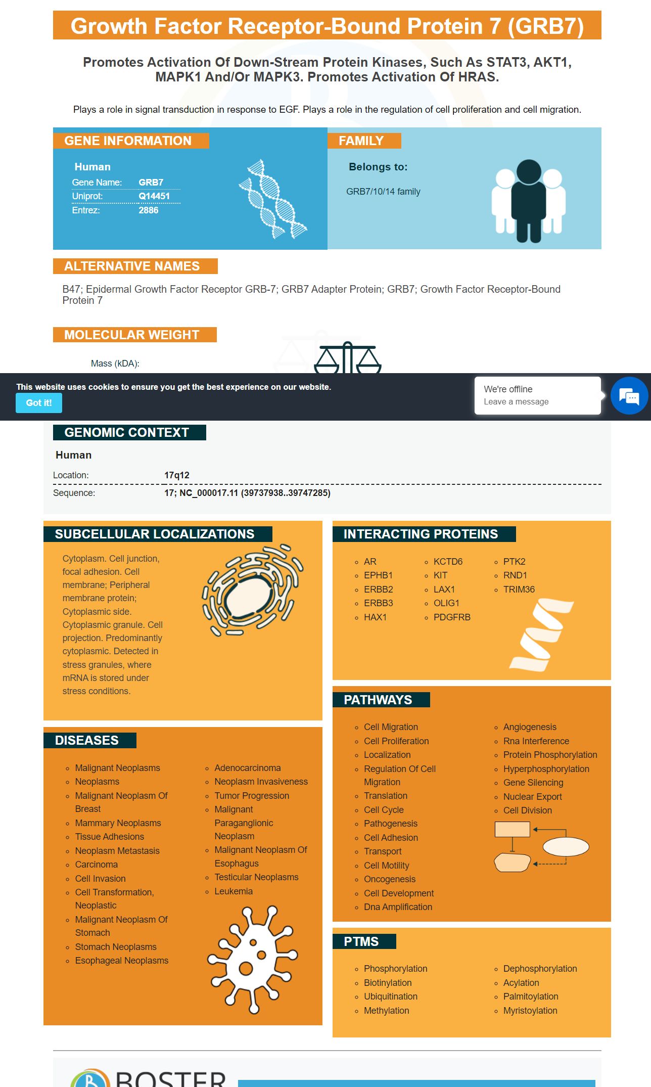
Plays a role in signal transduction in response to EGF. Plays a role in the regulation of cell proliferation and cell migration.
| Human | |
|---|---|
| Gene Name: | GRB7 |
| Uniprot: | Q14451 |
| Entrez: | 2886 |

| Belongs to: |
|---|
| GRB7/10/14 family |

B47; Epidermal growth factor receptor GRB-7; GRB7 adapter protein; GRB7; growth factor receptor-bound protein 7
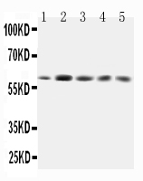
Mass (kDA):
59.681 kDA

| Human | |
|---|---|
| Location: | 17q12 |
| Sequence: | 17; NC_000017.11 (39737938..39747285) |
Cytoplasm. Cell junction, focal adhesion. Cell membrane; Peripheral membrane protein; Cytoplasmic side. Cytoplasmic granule. Cell projection. Predominantly cytoplasmic. Detected in stress granules, where mRNA is stored under stress conditions.


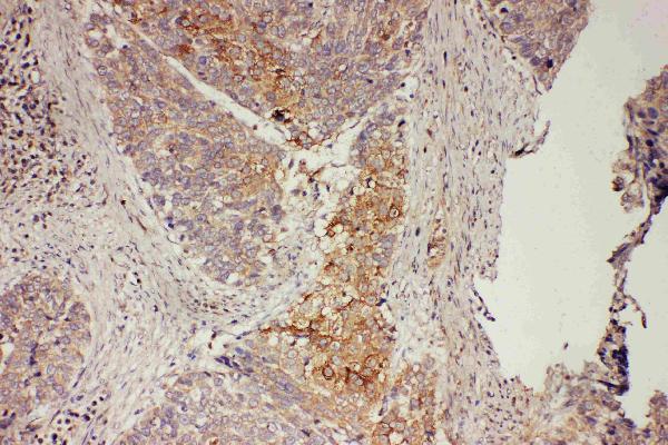
The best uses of boster proteins can be determined through IHC. It has been demonstrated in a human lung cancer cell line that boster proteins capture using a conventional chromatography step. Boster Bio's unique technology is applicable to scientists from all over the world. To learn more about boster bio's Biological applications, read on. Here are some best uses of boster proteins.
Polyclonal antibodies conjugated to GRB7 (a GRB7 gene) recognize antigen p21, a protein found in human colon cancer tissues. To optimize immunohistochemistry, antibodies must be specific for p21. To identify this antigen, a patient's tissue must be perfused to remove blood components and prevent reagent tainting.
The antibody must recognize the target antigen to create an image, but the process is multistep and requires optimization at every step. After identifying the target antigen, antibodies are diluted in buffer to stabilize the antibody and promote uniform diffusion into the sample. This reduces background staining. The buffers used for immunohistochemistry include normal serum, non-fat dry milk, bovine serum albumin, and gelatin. Common blocking buffers contain gentle surfactants that facilitate wetting.
The antibody polypeptide variants are designed to maintain the desired activity and similar binding affinity to the biomarker. Moreover, mutations must not result in reading-frame instability or create complementary regions, which would produce a secondary mRNA structure. The resulting IHC-optimized polyclonal antibodies using the GRB7 marker are the most appropriate choices for diagnosis and monitoring in clinical research.
The antibodies are designed for use in a variety of applications, including disease diagnosis and drug development. In cancer research, IHC can detect tumor markers in multiple tissues and monitor the activity of disease markers in patients. The antibodies also detect changes in the expression levels of the markers in the cells, and can be used in a variety of contexts, from drug discovery to basic research.
Biomarker-specific antibodies are typically screened on a polydoma. These polydomas are provided as multi-well tissue culture plates. In the initial antibody screening step, antibodies are selected that are specific for overexpression of MMP-26 in breast cancer tissues. The samples for these tests are normal breast tissue and stage I, II, and III tumors. To assess a biomarker's specificity, multiple antibodies may be added sequentially or simultaneously to a single sample.
ELISA kit for the detection of low-picogram levels of human connecting peptide is designed for research use. The peptide has a half-life three to four times that of insulin, and therefore serves as a useful measure of insulin production in beta cells. A home-made kit has lower costs, which make it a suitable choice for use in clinical settings. Its sensitivity is excellent, making it suitable for mass screening of people living in areas where fasciolasis is endemic.
The sensitivity of these immunoassays was further improved by the addition of gold nanoparticles to the test samples. Signal intensities were measured before and after the addition of gold nanoparticles. Blank measurements were also performed using PBS, which serves as a negative control. Signal intensities were divided by the number of blanks, and the ratios were calculated. The higher the ratio, the more sensitive the immunoassay is.
The GRB7 gene is located nearby the HER-2/erbB2 gene and is often co-amplified with it. Breast cancers often have high levels of GRB7 protein expression. While HER-2 overexpression alone has been shown to be an adverse prognostic factor, the presence of GRB7 protein is a more significant independent adverse risk factor. Biological applications of the GRB7 marker include the discovery that over-expression of GRB7 promotes HER-2-mediated signaling and facilitates tumor formation in an animal model.
Molecular analyses of GRB7 have shown that this gene is expressed in focal adhesions, the cytoplasm, and stress granules. A deletion of the SH2 domain abolishes GRB7 localization in focal contacts. The interaction between the PH domain and phospholipid recruits GRB7 to the cell membrane. It also plays a role in the nuclear-cytoplasmic export complex.
The GRB7 gene has been found to be expressed in normal human tissues, including breast and ovarian cancer. Research is ongoing to determine its clinical significance and how it may help to detect tumors. In breast and ovarian cancer, GRB7 expression has been shown to be correlated with tumor size, lymph node metastases, and overall survival. However, it is still unclear how the GRB7 gene may impact cancer treatments.
The expression of GRB7 is highly variable in different human tissues. The highest levels of GRB7 protein expression have been observed in the bone marrow, salivary glands, and brain, and it has a variable expression in skeletal muscle and the endometrium. The protein has almost no expression in the spleen and fallopian tubes. For these reasons, the GRB7 gene has great potential for use in research.
The GRB7 marker is a newly developed cytoplasmic protein that is overexpressed in many types of cancer, including gastric and esophageal. The marker is primarily expressed in the cytoplasm, where it interacts with upstream binding partners, such as members of the EGFR receptor family. Grb7 is also found in focal contacts, where it is phosphorylated by a protein called focal adhesion kinase. This protein is known to play a role in signal transduction and cell migration. The recruitment of Grb7 is a sign of Ras signaling.
The GRB7 gene is expressed in a variety of tissues and is characterized by several functional domains. The GRB7 gene product contains a unique proline-rich motif, Mig region, and SH2 domain. The C-terminal SH2 domain binds phosphotyrosine in the context of adjacent amino acids, and is critical for adapter function. Grb7 family members have also been shown to interact with tyrosine kinase receptors and proto-oncogenes.
Currently, Grb7 is a co-amplified gene with HER-2/neu in most breast cancer cell lines and primary breast tumors. There have also been associations between Grb7 and HER2/neu in western blot as well as in small-cohort studies. Furthermore, Grb7 is a marker of a subset of breast cancer patients with a decreased survival. Future studies of drug agents targeting Grb7 should include measurements of Grb7 levels in patients.
GRB7 antibody antibodies have been used to screen for a variety of tumors. Boster Bio also has a comprehensive line of antibody reagents for cancer research. Its Picokine(tm) platform allows high-sensitivity ELISA. These reagents have been validated and tested against a diverse panel of 250 tissues. Their sensitivity and specificity have been verified with picogram-level sensitivity.
PMID: 9125150 by Kishi T., et al. Molecular cloning of human GRB-7 co-amplified with CAB1 and c-ERBB-2 in primary gastric cancer.
PMID: 9710451 by Tanaka S., et al. A novel variant of human Grb7 is associated with invasive esophageal carcinoma.
*More publications can be found for each product on its corresponding product page