This website uses cookies to ensure you get the best experience on our website.
- Table of Contents


Facts about Glutaredoxin-3.

Required for hemoglobin maturation (PubMed:23615448). Does not possess any thyoredoxin activity since it lacks the conserved motif that is essential for catalytic activity.
| Human | |
|---|---|
| Gene Name: | GLRX3 |
| Uniprot: | O76003 |
| Entrez: | 10539 |

| Belongs to: |
|---|
| No superfamily |

FLJ11864; GLRX3; GLRX4; Glutaredoxin 3; glutaredoxin 4; glutaredoxin 5; GRX3; GRX4; PICOT; PICOTbA500G10.4; PKC-interacting cousin of thioredoxin; PKCq-interacting protein; thioredoxin-like 2; TXNL2; TXNL3
Mass (kDA):
37.432 kDA

| Human | |
|---|---|
| Location: | 10q26.3 |
| Sequence: | 10; NC_000010.11 (130136375..130182877) |
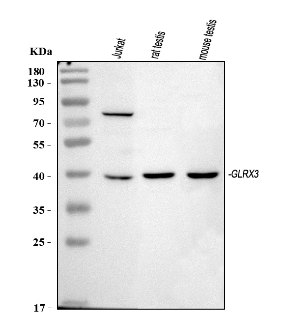
Expressed in heart, spleen, testis and, to a lower extent, in thymus and peripheral blood leukocytes. Weakly expressed in lung, placenta, colon and small intestine.
Cytoplasm, cytosol. Cytoplasm, cell cortex. Cytoplasm, myofibril, sarcomere, Z line. Under the plasma membrane (By similarity). After PMA stimulation, GLRX3 and PRKCQ/PKC-theta translocate to a more extended submembrane area (By similarity). In the Z line, found associated with CSRP3 (By similarity).


The GLRX3 marker is a protein expressed in E.coli that is suitable for SDS-PAGE. It is not endotoxin labeled and can be kept at 2-8°C for one week. Glutaredoxin 3 can be used in conjunction with all Boster products. You can use it to analyze the concentration of protein in different samples, such as for in vitro testing and downstream applications.
Boster Bio has a membrane staining kit that allows researchers to assess the efficiency in protein transfer from polyacrylamide gelatines to membranes. A total protein stain can be used to identify proteins not transferred to the membrane. They can also confirm the transfer efficiency using irregular bands or no bands. The protein transfer efficiency can also be analyzed by monitoring the band intensity in each lane.
Boster Bio membrane solution is a complete lysis buffer that can be used with many immunoassay and protein purification applications. This buffer does not affect the immunoreactivity of proteins or degrade them. Its non-ionic nature allows it to be used in Western Blot. Stripping Buffer is another Boster product that helps to remove primary antibodies from probed cells. This solution removes both primary and secondary antibodies without affecting immobilized antigen. It allows researchers to reuse the membrane and avoid cross-contamination.
Tests for transfer efficiency were conducted using pooled samples of human serum. These were separated with 10% SDS–PAGE and transferred onto PVDF or nitrocellulose membranes. The membranes were fixed with acetone or 50% methanol. The samples then were stained with LCA and SNA. After the fixation procedure, the membranes were stained with 50% methanol/water. The exposure time for each experiment was identical to the traditional fixation process.
The cell surface must first be treated with an autoradiography film to determine the transfer efficiency. This film needs to be exposed for approximately 5 minutes. This film also requires darkroom. A WB Developing Fixing kit can be used to develop an autoradiography film. You can also place a thin layer X-ray film over the membrane, and let it develop for 10 minutes. The amount and duration of the antigen bind to the membrane will dictate the exposure time. Multiple exposures may be necessary to achieve the desired signal-to-noise ratio.
AAL and PHA-E staining were performed on serum samples from prostate cancer patients and healthy volunteers. After transferring to the gel, the proteins were detected by using a chemoluminescent substrate. Using this method, researchers can evaluate the efficiency of protein transfer from a protein-staining gel. They can also perform a Boster Bio test to determine the protein concentration in a sample containing multiple antibodies.
GLRX3 Marker regulates c–MET, PI3K and AKT signaling pathways. Its expression alters epithelial-mesenchymal transition and cancer stemness. In a recent study, researchers measured the serum level of GLRX3 in patients with PDAC compared to healthy controls. Results showed that serum GLRX3 levels correlate with shorter disease-free survival and overall survival (OS). These differences were not statistically significant.
The GLRX3Marker, a non-religious marker protein, is found in cells. The protein has three domains. Its presence in cells can help determine if they have cancer. It can also detect pancreatic tumors. Studies have shown GLRX3 could help diagnose pancreatic Cancer in people with this disease. Its main applications are:
GLRX3 can be described as a non-recombinant proteins with an N terminal thioredoxin like domain and two domains PICOT homology. It is widely distributed and has been found in many tissues. It is expressed in epithelial cell epithelium, but its function remains unclear. It has been linked with cardiac contractility, and PKC-theta regulation.
GLRX3 knockdown mice cells induced Ecadherin/vimentin, two epithelial markers that EMT, in mice. GLRX3 knockdown cells had lower expression of Wnt pathway-related protein. These results suggest that GLRX3 knockdown cell lines are more likely to have pancreatic cancer-like features and are more susceptible to metastasis.
GLRX3 antibodies were used at a 1:1000 concentration in various cell lines to perform the immunoblot analysis. Primary antibodies were anti-human GLRX3 and beta-catenins, E-cadherins, GAPDH and ABCG2. These results were confirmed by duplicates of each experiment in triplicate. GLRX3 knockdowns cells also detected Wnt1, 5a and c–MET.
GLRX3 proteins were detected in pancreatic tumor cell lines. GLRX3 protein expression was 8.8 times higher in pancreatic carcinoma than in healthy tissue. HPDE cells, on the other hand, only a small percentage expressed GLRX3.
ECL chemistry, a common chemistry, is used to detect proteins expression in cell cultures and in many other applications. This technology uses luminol-based chemiluminescent compounds to recognize a variety of target proteins. For example, Boster Bio's ECL Western Blotting Substrate detects horseradish peroxidase and is compatible with nitrocellulose membranes and various blocking buffers. Boster Bio's Western Blotting Substrate features low background levels, and can be visualized with x-ray film or CCD imaging systems.
ECL is not a perfect technology, as its detection sensitivity is low and it is not possible to detect small amounts of target protein. Researchers can still use ECL to analyze small amounts and accurately detect their concentration. The ECL chemiluminescent detector system has a strong signal that can be used to reimagine samples without losing its intensity.
The C-DiGit(r), Blot Scanner is a great choice for those who don't want to spend money on darkrooms or develop reagents. The system has a wider dynamic spectrum than film, so it can detect target proteins in fewer exposures. It is also easy-to use and has long shelf life. The iBright Imaging System can be used for analysis of protein expression in cell culture samples, and other experiments.
Amersham ECL Prime chemiluminescent detection tool uses Amersham ECL Prime. Multiple blots can run at once. The ECL-reagent is extremely sensitive. It can detect protein levels at an average of 23 kDa. Hybond ECL membranes have a 0.45 pores size and are highly absorbent nitrocellulose membranes.
Clarity Max substrates will work with any horseradish/peroxidase conjugate. This makes them an excellent choice in western blots. The Clarity Max can be used with all western blotting applications that have abundant proteins. The Clarity Max substrates for chemiluminescence are compatible to any horseradish Peroxidase conjugate.
DAB chromogenic analysis is a powerful method of cell and tissue cellular analysis. DAB chromogen stain detects apoptotic colonic epithelial cells. The method uses a Methyl Green counterstain, which is made up of 0.125% VWR brand powder in 0.1M sodium acetate buffer at a pH of 4.2-4.3. Tissue samples were mounted onto slides the previous night and subjected to a series of ethanol drying steps. Methyl Green (more nuclear in the cortex) is used for detection.
PMID: 10636891 by Witte S., et al. Inhibition of the c-Jun N-terminal kinase/AP-1 and NF-kappaB pathways by PICOT, a novel protein kinase C-interacting protein with a thioredoxin homology domain.
PMID: 11124703 by Stanchi F., et al. Characterization of 16 novel human genes showing high similarity to yeast sequences.