This website uses cookies to ensure you get the best experience on our website.
- Table of Contents
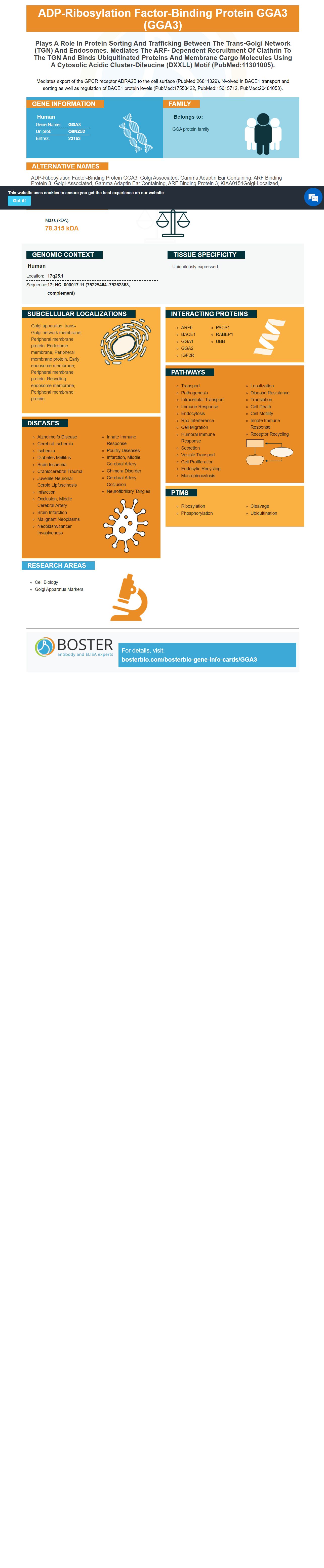
Facts about ADP-ribosylation factor-binding protein GGA3.
Mediates export of the GPCR receptor ADRA2B to the cell surface (PubMed:26811329). Nvolved in BACE1 transport and sorting as well as regulation of BACE1 protein levels (PubMed:17553422, PubMed:15615712, PubMed:20484053).
Human | |
---|---|
Gene Name: | GGA3 |
Uniprot: | Q9NZ52 |
Entrez: | 23163 |
Belongs to: |
---|
GGA protein family |
ADP-ribosylation factor-binding protein GGA3; golgi associated, gamma adaptin ear containing, ARF binding protein 3; golgi-associated, gamma adaptin ear containing, ARF binding protein 3; KIAA0154Golgi-localized, gamma ear-containing, ARF-binding protein 3
Mass (kDA):
78.315 kDA
Human | |
---|---|
Location: | 17q25.1 |
Sequence: | 17; NC_000017.11 (75225464..75262363, complement) |
Ubiquitously expressed.
Golgi apparatus, trans-Golgi network membrane; Peripheral membrane protein. Endosome membrane; Peripheral membrane protein. Early endosome membrane; Peripheral membrane protein. Recycling endosome membrane; Peripheral membrane protein.
Boster Bio Anti GGA3 marker detects GGA3 across a wide range cell types. Validated for IHC, WB and Immunofluorescence. Boster is committed to providing a high quality product for every lab. In this review, we will discuss the advantages and features of Boster Bio Anti-GGA3 Marker.
Boster bio Anti-GGA3 marker antibodies are monoclonal anti-bodies that react with Humans as well as other species. They are nonhazardous and are available without BSA. You can also purchase blocking peptides at 1.0 mg/ml, with the cost depending on the length of the immunogen. These antibodies are tested on positive and negative samples to ensure that they are reactivity. Always use a high quality primary antibody for the best results.
GGA3 can also be detected in epithelial cells, brain, and other cell types. The VHS domain, which is part of the protein's membrane proteins in Golgi, is involved in protein transport at the TGN. However, this protein has also been shown co-localizes in peripheral cytoplasm with AP-1 & AP-3.
Researchers examined the intracellular localizations of GGA proteins in human cells using immunoelectron microscopes. Endogenous GGA3 exhibited very little signal. However, transfection with higher levels of untagged GGA3 resulted specifically labeling the Golgi-stack region. The labeled regions had gold particles that were marked by a budding membrane profile, and an electron-dense coat. Double-labeling the regions with anti-TGN46 antibodies further confirmed the presence TGN.
ORFs were identified by PCR, 5'RACEPCR and PCR in order to isolate both the short and long GGA3 genes. Primers were designed using the sequence of DNA clone KIAA0154 (GenBank accession number D63876). The PCR product was then subcloned to pCR3.1 vector. This vector was designed for expression GST fusion protein.
The protein coat assembly process involves the coordination of many regulatory or structural proteins. The coat assembly proteins form dynamic networks with low affinity interactions, which lead to stable coats on membranes. GGAs are modular and have a VHS region and GAT regions linked by a linker section. GGA3 is colocalized with Golgi markers, and can be found on coated vesicles or buds in the TGN. Its dissociation can be caused by overexpression of the ARF1 gene.
It is known that GGA and TCS proteins interact. GGA3 was able to identify the DXXLL motif using GST-pulldown assays. Using GST-TCS conjugated glutathione agarose beads, the mutants were incubated with HepG2 cell lysates and transfected separately with indicated plasmids. Both proteins were bound by GST-TCS proteins, which were then quantitively analyzed.
GST-GGA3GAT is a fluorescent protein that specifically binds to GGA3 in a nucleotide-dependent manner. GST-GGA3GAT binds to the GAT domain of the human effector protein GGA3. As such, it has a role in ARF1-mediated deposition of protein coats on membranes. GGA3 activation status may also be determined by GAT.
CI-MPR is a transmembrane glycoprotein that delivers newly synthesized hydrolases from the TGN to pre-lysosomal compartments. It is found in approximately 10-20% the cell membrane. GGA proteins bind TCS, which alters their interaction. To test this, we used an anti-GGA3-antibody in HepG2 cell-lysates with increasing TCS load.
Validation of antibodies can be difficult. This method requires the use of purified protein as an immunogen. Antibodies that are in their native form are more effective than those that have been denatured. ENCODE must accept antibodies that recognize over 10% of the bound peak to approve them. The antibody must be validated on WB if it is to be accepted by ENCODE. It should show a signal of 50% or more over other bands, or a unmodified recombinant histone.
Companies with the highest level of validation do not disclose the specific details of their validation process. They should also provide an appropriate protocol and dilutions for every application. These steps may be simple and cost-effective, but they may not suffice for all applications. Some antibodies may not show promising results if validated on WB and IHC. To avoid these problems, make sure you read the validation sheet before you use the antibody.
Another important criterion when validating antibodies is reproducibility. This criterion identifies whether the antibody is reproducible against another validated antibodies or a different method of measuring the target. While this is not generally possible with antibodies there are a few exceptions. For example, an antibody used in the analysis of HER2 in a tumor cell line was not reproducible.
Validation of antibodies are crucial for accurate WB/IHC analyses. Validated antibodies are essential for reproducible and accurate results. Although validation of antibodies can be complicated, they can be used for immunofluorescence analysis. Researchers can use online portals such as Antibodypedia to access data. This portal accepts commercially available antibodies and original experimental data.
It is important that you note that a product may be validated on WB by a qualified pathologist, laboratorian, and IHC by an independent laboratory. The manufacturer must be able to provide an appropriate quality control sylicm. The quality control materials should be easily accessible. To ensure reproducibility, manufacturers should provide information on the tissue used to stain. This information should be presented in a user-friendly format.
To ensure validity, the study samples must be collected according to the correct criteria. Additionally statistical controls should also be performed to eliminate selection bias. The samples must be drawn from clinically relevant samples. WB or IHC studies will not be able to support prognostication and quantitative claims. Immunofluorescence is also highly sensitive in detecting antibodies.
To ensure the accuracy of the antibodies, the manufacturer should conduct titer test on paraffin embedded cell pellets, cell-line pellets and patient tissues. A good antibody should not only stain target cells but also decrease with dilution. It should also be consistent with published literature in terms of expression patterns. There are also many advantages to using antibodies that have been validated on WB, IHC, and Immunofluorescence.
PMID: 10747089 by Dell'Angelica E.C., et al. GGAs: a family of ADP ribosylation factor-binding proteins related to adaptors and associated with the Golgi complex.
PMID: 10749927 by Boman A.L., et al. A family of ADP-ribosylation factor effectors that can alter transport through the trans-Golgi.
*More publications can be found for each product on its corresponding product page