This website uses cookies to ensure you get the best experience on our website.
- Table of Contents
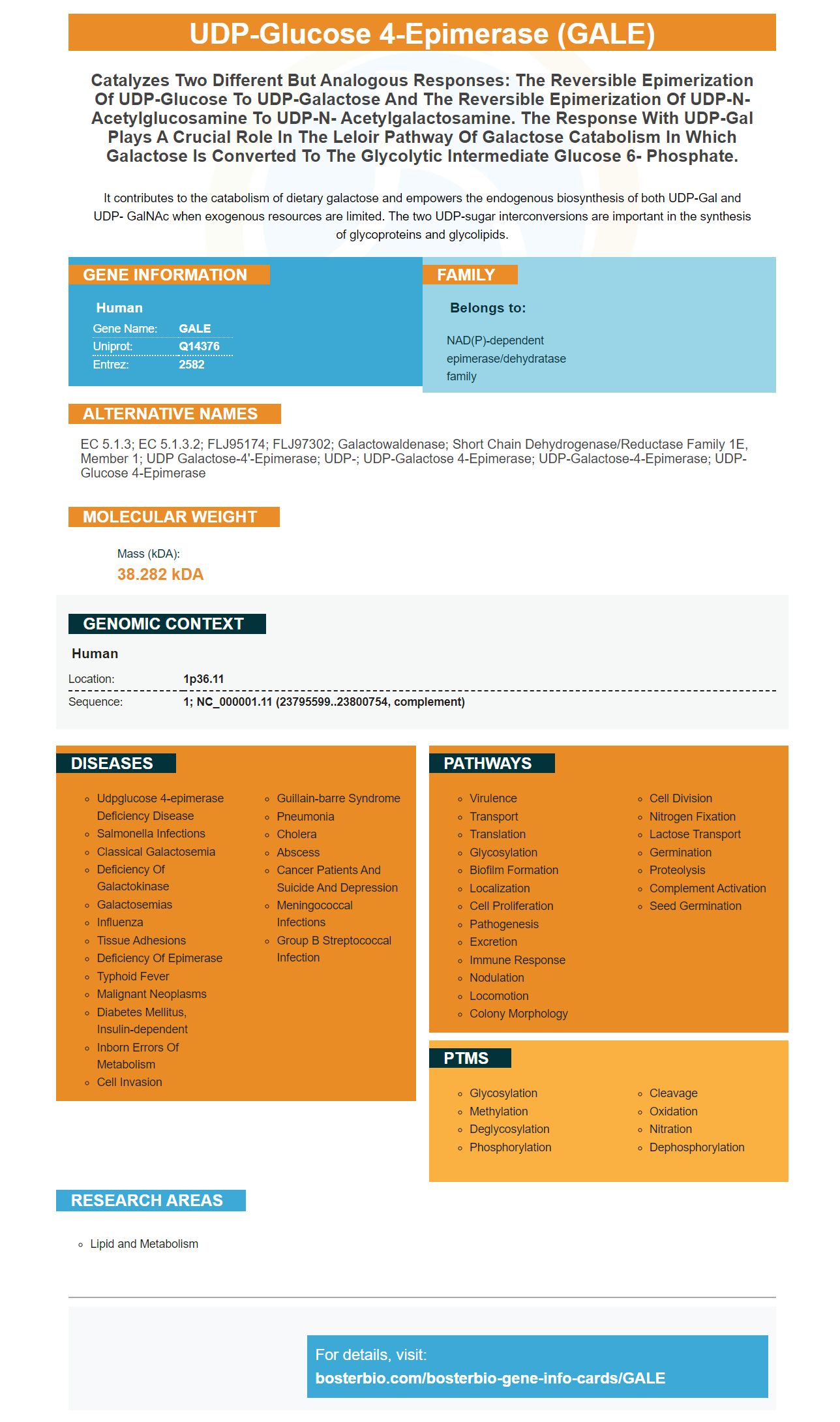
Facts about UDP-glucose 4-epimerase.
It contributes to the catabolism of dietary galactose and empowers the endogenous biosynthesis of both UDP-Gal and UDP- GalNAc when exogenous resources are limited. The two UDP-sugar interconversions are important in the synthesis of glycoproteins and glycolipids.
Human | |
---|---|
Gene Name: | GALE |
Uniprot: | Q14376 |
Entrez: | 2582 |
Belongs to: |
---|
NAD(P)-dependent epimerase/dehydratase family |
EC 5.1.3; EC 5.1.3.2; FLJ95174; FLJ97302; Galactowaldenase; short chain dehydrogenase/reductase family 1E, member 1; UDP galactose-4'-epimerase; UDP-; UDP-galactose 4-epimerase; UDP-galactose-4-epimerase; UDP-glucose 4-epimerase
Mass (kDA):
38.282 kDA
Human | |
---|---|
Location: | 1p36.11 |
Sequence: | 1; NC_000001.11 (23795599..23800754, complement) |
Optimizing your experiments for ELISA requires you to be aware of several things. First, you must understand that ELISA requires several choices at almost every step. You must choose the blocking buffer, antibody level, and sample preparation. Boster Bio provides optimization tips and guidelines to help you answer these questions. These guides can also be used to optimize your experiments. These guides will also help you optimize your experiments.
The first step in quantitative analysis of Western blots is to define the lanes to analyze. The lane frames must be defined so that they include all signals from each band within each lanes. Once you have defined the lane outlines, it is possible to create a profile. This tool will normalize your data so you can infer whether your protein level is truly up-regulated or down.
Normalization is a method used to reduce the effects of experimental error. Pipetting errors, sample concentration errors, or protein transfer errors can all lead to experimental error. The process of normalization provides confidence that the signal differences reflect true protein expression levels and corrects for these errors. You can make comparisons more meaningful and ensure accuracy with your analysis by using a normalization reference. Here's how the process works.
A western blot is able to analyze protein expression levels using two types of agarose Gel. The first type, the stacking, is slightly acidic with a low acrylamide content. This allows the gel to have a more defined band. The second type, also known as the separating gel is more basic and contains more polyacrylamide. This gel narrows pores, making it easier for smaller proteins to travel across it.
Different types can be used for detection. There are many ways to detect the level of protein expression using western blot. For example, you can use either a fluorescent or a radioisotope-conjugated primary antibody. The difference is that the radioisotope-conjugated primary antibody uses an enzyme and fluorophores, while the former uses proteins to label.
There are other ways that housekeeping proteins can be reduced. Another method, known as total protein normalization allows you to determine total levels of protein by staining the membrane with specific colors. Bio-Rad's Stain-Free technology is the best. Researchers use GALE markers to validate proteins within complex biological samples. This technique has many advantages, including its sensitivity and linear dynamic range.
The GALE marker should not be used in excess. You should use a standard dilution to reduce the amount of primary antibodies. In some cases the primary antibodies might not be sufficient to detect a protein on the membrane. Antibodies against phosphorylated proteins, for example, may have a higher affinity than antibodies against non-reactive proteins. It is important to experiment to determine the best buffer.
The procedure for analyzing the protein expression levels was performed on 8 cell lines. Diagenode Blue ladder HRP antibody (Cat. No. C15200202 was used to detect alpha tubulin protein. The antibody was dispersed in TBS/Tween containing 5% skimmed Milk at a 1:1,000 dilution. The band quantity can be seen on the right using a marker in kD.
The Gale Marker in the Martian landscape is a singular feature. It was created by changes in rock formation. This formation could be explained by three possibilities. One scenario suggests that cementation was caused when a layer containing water that was chemically abundant was present. The second scenario is that the cementation occurred because of sulfate rich sediments.
Below is an image that shows the marker's horizon and its relationship with the nearby cone. The marker is located on the horizon of the Context Camera (CTX) mosaic. Each subset contains one photo that shows the marker horizon. These images were enhanced for its location. Each image shows 50m. The different stretch applied to each image affects its colors. The images were acquired by the Curiosity rover, which has driven across the Gale crater's sediments and measured the rock's properties in detail.
GALE markers can be used in IHC to identify specific target molecules. Boster Bio offers a variety of secondary antibodies, isotype controls, and detection systems optimized for IHC. We answer several customer questions about negative controls. Continue reading to learn how to create the perfect conditions for negative control. Alternatively, check out our guide to GALE marker optimization.
PMID: 8593531 by Daude N., et al. Molecular cloning, characterization, and mapping of a full-length cDNA encoding human UDP-galactose 4'-epimerase.
PMID: 9538513 by Maceratesi P., et al. Human UDP-galactose 4'epimerase (GALE) gene and identification of five missense mutations in patients with epimerase deficiency galactosemia.