This website uses cookies to ensure you get the best experience on our website.
- Table of Contents
1 Citations
2 Citations 11 Q&As

3 Citations 6 Q&As

2 Citations 3 Q&As


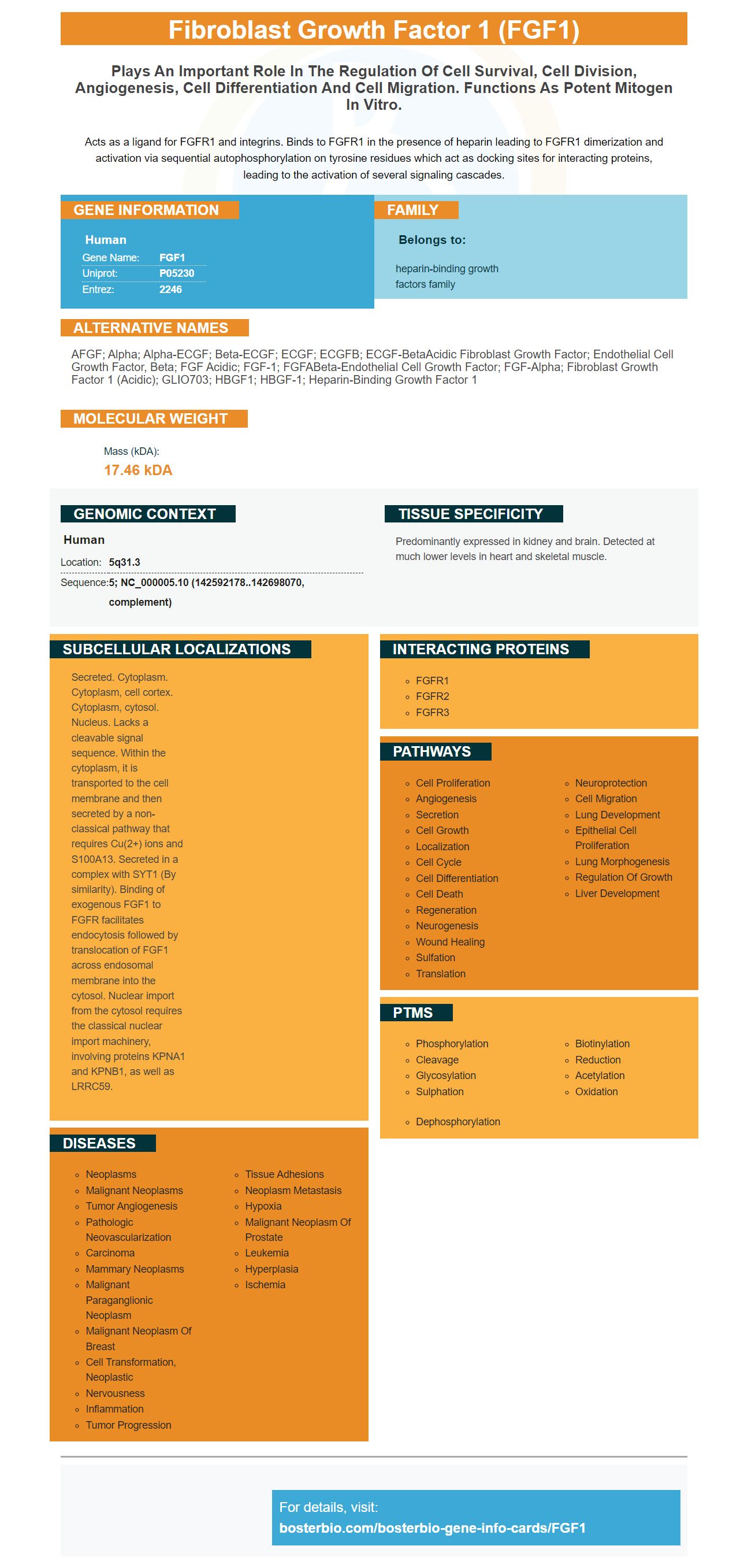
Facts about Fibroblast growth factor 1.

Acts as a ligand for FGFR1 and integrins. Binds to FGFR1 in the presence of heparin leading to FGFR1 dimerization and activation via sequential autophosphorylation on tyrosine residues which act as docking sites for interacting proteins, leading to the activation of several signaling cascades.
| Human | |
|---|---|
| Gene Name: | FGF1 |
| Uniprot: | P05230 |
| Entrez: | 2246 |

| Belongs to: |
|---|
| heparin-binding growth factors family |

AFGF; alpha; alpha-ECGF; beta-ECGF; ECGF; ECGFB; ECGF-betaAcidic fibroblast growth factor; endothelial cell growth factor, beta; FGF acidic; FGF-1; FGFABeta-endothelial cell growth factor; FGF-alpha; fibroblast growth factor 1 (acidic); GLIO703; HBGF1; HBGF-1; heparin-binding growth factor 1
Mass (kDA):
17.46 kDA

| Human | |
|---|---|
| Location: | 5q31.3 |
| Sequence: | 5; NC_000005.10 (142592178..142698070, complement) |
Predominantly expressed in kidney and brain. Detected at much lower levels in heart and skeletal muscle.
Secreted. Cytoplasm. Cytoplasm, cell cortex. Cytoplasm, cytosol. Nucleus. Lacks a cleavable signal sequence. Within the cytoplasm, it is transported to the cell membrane and then secreted by a non-classical pathway that requires Cu(2+) ions and S100A13. Secreted in a complex with SYT1 (By similarity). Binding of exogenous FGF1 to FGFR facilitates endocytosis followed by translocation of FGF1 across endosomal membrane into the cytosol. Nuclear import from the cytosol requires the classical nuclear import machinery, involving proteins KPNA1 and KPNB1, as well as LRRC59.


You may be wondering what the most effective applications of the FGF1 marker are. This article will discuss the many functions of the FGF1 marker, which includes the development of biomaterials the neuroprotective properties of the marker, and the treatment of cancer. For more information on Boster Bio, please contact Sanbio. We're pleased and able to assist you with your research. You can also purchase customized services as well as BeNeLux delivery.
In vitro angiogenesis is measured through the tube formation assay. Huang et al. performed this assay using OGD-treated HBMECs. Matrigel was applied to each well and incubated for 30 minutes to set. The treated cells were then seeded in the plate and incubated for 16 hours at 37°C. The cells were then counted using a bright field microscope.
Boster Bio has a commercially available anti FGF1 antibody that can be utilized in a variety of applications. Their FGF1 antibody reacts to Human. It can be stored at -20°C or 4degC. You can also purchase the antibody as a blocking protein separately, depending on its length. For more details, read Boster Bio's FGF1 antibody review.
Angiogenesis is a key function for the FGF1 marker. When administered, nmFGF1 encourages the development of angiogenesis. It also activates the S1P1 signaling pathway. This pathway is activated after stroke. In a recent study Boster Bio and the University of Minnesota published the results of their clinical research on this marker. This research suggests FGF1 may be a useful treatment for ischemic stroke.
Adult stem cells are essential for tissue homeostasis and can also be utilized as regenerative agents. They self-renew. At at least one stem cell is retained in every cell division. Only after symmetric division does the pool shrink due to cellular decline. Numerous studies have demonstrated that FGF signaling is an essential regulator of tissue healing.
The FGF1 molecules is a multipotent growth factor that promotes proliferation, differentiation, and survival of a wide range of cells. Studies have shown that ADMSCsFGF1 conditioning medium can increase the growth of fibroblast 3T3 cells from the NIH. FGF1 also plays a role in angiogenesis and is more potent than vascular endothelial growth factor (VEGF).
The process of aging is an underlying process that causes the degeneration of many organs. It is characterized by high metabolic demands as well as mitotic activity, and regular regeneration. In the process, many tissues lose their regenerative capabilities. Recent research has demonstrated that adult stem cells can be found in almost every organ. They are able to sustain tissue homeostasis, repair, growth and maintenance. These cells can help to regenerate tissue and treat disease and are a powerful source of stem cells for many therapeutic applications.
During embryonic development, neural stem cells arise from the neuroepithelium. They are initially multipotent, but later in their development, they are limited to a specific area like the cerebellum and the subgranular part of the hippocampus. The FGF receptors, FGFR-1 and FGFR4 are present in adult mESCs.
It is unclear what function FGF1 plays in the regeneration of nerve tissue and neuroprotection. The hormone has been linked to nerve regeneration and healing after injury. Exogenous FGF1 can rescue neurons and promote regeneration of sensory axons after dorsal root segregation. Additionally, subcutaneous FGF1 injection delayed senescence and preserved cholinergic cells in the medial septum.
The FGF1 immunoreactivity in the hypoglossal nucleus was comparable in both the rostral and caudal regions. Neurons that are immunoreactive with FGF1 showed clear staining in their cytoplasm and neural processes. FGF1 immunoreactivity in DMNV was seen in small neurons as well as the hypoglossal nuclear nucleus. Medium-sized oval-shaped, 30-50 um in diameter neurons were in the presence of FGF1. FGF1 was detected within the neural processes' cytoplasm and neurons within the facial nucleus.
In an animal model, the FGF1 and PSD95 interactions are altered. In addition, FGF-2 neopterin, and methamphetamine dependence modify the FGF-GF1-GFP relationship. Different recursive partitioning models can result in different thresholds for FGF-1. The complex interaction between FGF1/FGF-2 as well as neurocognitive functions is a consequence of.
Studies on mice reveal that FGF1 is expressed in cholinergic nerves, including the hypoglossal and thalamus nucleus. It is also found in the lateral region of the sulcus motor nucleus that is located in the dorsal region of the vagus and facial nucleus. Although FGF1 and ChAT both co-localize in DMNV, FGF1 immunoreactivity was highest in the rostral and lateral pons.
Boster Bio offers a wide variety of antibodies that detect this tumor-associated protein. These antibodies are highly specific, sensitive, and can be used in Western blotting and IHC. Boster Bio antibodies are based on new technology, including picogram-sensitive ELISA. The company has been manufacturing antibodies since 1993 and has spent more than two decades of experience to develop their techniques. Boster Bio antibodies have been featured in more than 29,000 scientific publications. Boster Bio antibodies have been validated for WB, IHC, and Cytometry. Cytometry.
Numerous tumors with high levels of FGF1 receptor have been discovered by researchers. This makes it an attractive molecular target for treating cancer. Cytotoxic conjugates, which target cancer-specific FGFR1 are able to enter tumor cells through receptor-mediated endocytosis. The effectiveness of the internalization process is contingent on how the receptor is dispersed within the plasma membrane. The ability to cluster the receptor into larger oligomers boosts its uptake and allows for multiple endocytic routes.
Additionally, miR-144 is a tumor-specific RNAi cell is an essential component in the proliferation of tumor-related cells and metastasis. It also plays a significant role in cancer cell proliferation and drug delivery, bypassing healthy cells. As more research is done to better understand its mechanisms the anti-cancer function of miR-144 becomes more apparent.
Prostaglandins and various angiogenic proteins are essential to the process of angiogenesis. Angiopoietin-1 inhibits leukocyte adhesion towards endothelial cells , and decreases E-selectin, VCAM-1 expression. However, the precise mechanisms that cause angiogenesis remain unknown. Certain compounds may hinder angiogenesis as well as trigger the growth of new blood vessels.
Different growth factors are released by tumor cells during the process of tumor development. These factors may trigger the growth of capillaries in the tumor. The new blood vessels supply nutrients to the tumor. The blood vessels are dilapid and irregular in their shape. While some doctors view angiogenesis as is a path to waste but other doctors consider it essential for the growth of tumors. Angiogenesis inhibition is a useful therapeutic option.
Sprouts move towards the source of the angiogenic stimulation. Endothelial cells are able to migrate in tandem, forming loops and eventually the vessel lumen. The spitting process occurs at a rate of millimeters per day. The process allows new blood vessels to grow through the gaps in the vasculature. However, this process differs from splitting angiogenesis which involves the formation of new blood vessels.
Angiogenesis is a complicated biological process that involves different cells that play different roles. The FGF system can regulate other growth factors and may be upstream to specialized growth factor systems, such as VEGF , which is a cell-specific endothelial growth factor, PDGF (for smooth muscle cells). The figure illustrates the indirect angiogenic process. This study may be interesting to you If you're interested in finding out more about the angiogenic process.
This study investigated the role played by FGF1 in wound healing. Researchers used 40 mice of different age groups to assess the rates of wound healing for the two groups. A semi-quantitative scale was used to measure the healing process. The results indicate that the FGF1 marker contributes to wound healing. Further research is needed to determine the role of FGF-related pathways to heal wounds.
The normal biologic function of FGFs is believed to be involved in wound healing and the possibility of therapeutic intervention is being investigated. FGFs regulate cell proliferation and differentiation, as well as the response to injury. This includes wound healing. They also play a role in tumorigenesis. Twenty-two FGFs are known and have 13-71% amino acids identity. Each one is therapeutic and is involved in wound healing.
The regulation of FGF-10's mRNA is still a subject of debate. Beer et al. concluded that FGF-10's mRNA was not produced during wound healing in human skin. However, they did find that the protein form was stored within the wound and released following injury. In another study, Tagashira et al. showed that the expression of FGF-10's mRNA was increased within a day of an injury, and then returned to normal on the third day. Further studies have demonstrated that the application of FGF-10 to the skin resulted in wound healing.
In vivo, BP1 expression speeds wound healing. Additionally, it increased the angiogenic sprouting of animals. FGF-receptor-kinase inhibitors prevented its effects. The effects of BP1 were not significant, however the results indicate that FGF-1 enhances wound healing in adult animals. This research suggests the possibility of combining existing treatments with novel approaches.
PMID: 3523756 by Jaye M., et al. Human endothelial cell growth factor: cloning, nucleotide sequence, and chromosome localization.
PMID: 2590193 by Mergia A., et al. Structural analysis of the gene for human acidic fibroblast growth factor.
*More publications can be found for each product on its corresponding product page


