This website uses cookies to ensure you get the best experience on our website.
- Table of Contents
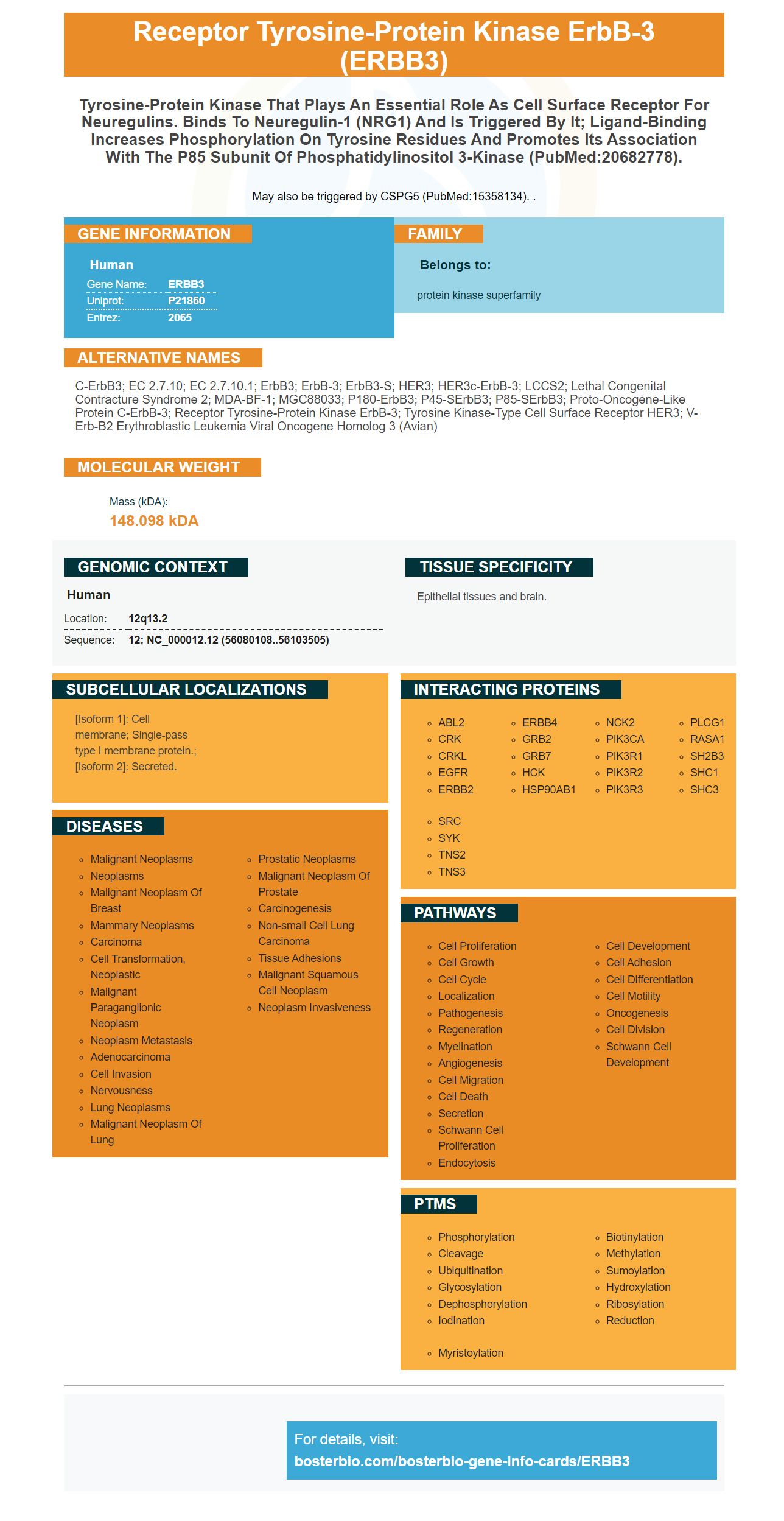


Facts about Receptor tyrosine-protein kinase erbB-3.

May also be triggered by CSPG5 (PubMed:15358134). .
| Human | |
|---|---|
| Gene Name: | ERBB3 |
| Uniprot: | P21860 |
| Entrez: | 2065 |

| Belongs to: |
|---|
| protein kinase superfamily |

c-erbB3; EC 2.7.10; EC 2.7.10.1; ErbB3; ErbB-3; erbB3-S; HER3; HER3c-erbB-3; LCCS2; lethal congenital contracture syndrome 2; MDA-BF-1; MGC88033; p180-ErbB3; p45-sErbB3; p85-sErbB3; Proto-oncogene-like protein c-ErbB-3; receptor tyrosine-protein kinase erbB-3; Tyrosine kinase-type cell surface receptor HER3; v-erb-b2 erythroblastic leukemia viral oncogene homolog 3 (avian)
Mass (kDA):
148.098 kDA

| Human | |
|---|---|
| Location: | 12q13.2 |
| Sequence: | 12; NC_000012.12 (56080108..56103505) |
Epithelial tissues and brain.
[Isoform 1]: Cell membrane; Single-pass type I membrane protein.; [Isoform 2]: Secreted.


The ERBB3 proteins plays a key role in the regulation a number of hormones, including EGFR. EGFR may be activated by a variety ligands. A monoclonal anti-ERBB3 antibody may be able provide effective therapy for many diseases. Scientists are currently trying to understand if the ERBB3 gene activates EGFR. The ERBB ligands activate the EGFR and are known as ERBB ligands.
The ERBB proteins regulate cellular growth, migration, and survival. Many human malignancies are linked to mutations in or overexpressions of ErbB proteins. Overexpression of the HER2 gene is seen in breast and colorectal carcinomas. These types of cancers often have poor prognoses. Boster Bio ERBB3 ligands target and inhibit ErbB3 signaling.
Experimentally determined total amounts ERBB allowed for the calculation of ERBB phosphorylation dynamic. All cell types were tested for the same reaction rates. Five minutes following ligand stimulation, ERBB phosphorylation levels were measured. These data were compared to global models. Boster Bio developed a proprietary method to calculate phosphorylation levels for the ERBB receptor in human cell.
Fig. 6. These models show that the ratio between phosphorylation rate constants and ERBB phosphorylation dynamics contributes to the diversity. Similar results were seen in the D-symmetric and unsymmetric models. The P-symmetric model shows a significantly smaller response number than the PD-symmetric model.
ERBB3 ligands bind with CRART16 in cytoplasm and increase ERBB3 expression. Cetuximab resistance was caused by CRART16 excess expression. CRART16 was overexpressed, which resulted in cetuximab resistant cells. However, ERBB3 overexpression enhanced the stemness properties CRC cells. These results demonstrate the therapeutic potential of cetuximab resistant cells.
For analysis, gene transfer plasmids containing a gene sequence of ERBB2 & ERBB3 were used. ERBB3 was highly expressed in MCF7 cell lines. In A431 cells, ERBB2 was expressed in higher amounts than ERBB1.
ERBB ligands activate EGFR in an interaction with the tyrosine kinasedomain. The phosphorylation of intracellular signaling cascades is then initiated by phosphorylation. The phosphorylated tyrosine residues on the EGFR bind intracellular signaling molecules, such as phosphoinositide-3-kinase. This is called negative cooperativity. Moreover, the tyrosine kinase domain is heterogeneous and dynamic, with different affinities to partner molecules.
The ERBB ligands bind directly to the EGFR’s intracellular juxtamembranedomain. There, a three part nuclear localization sequence mediates emergence of a fraction (or less) of the full-sized EGFR receptor in the nucleus. Similar signalling pathways are found in ErbB molecules, it is interesting. This trafficking is enabled by importin-b1. The nuclear pore complex also plays a role when EGFR is transported from the plasma membrane to the nucleus. Finally, the presence or absence of intranuclear intranuclear GFR is indicated by an active nuclear export signal within the nucleus.
Monoclonal monoclonal antibodies against EGFR have gone through multiple phases in clinical trials. However, FDA approval for broad-scale use has yet to be granted. These anti-EGFR medications have not shown any improvement in clinical outcomes. These findings indicate that ERBB ligands, a novel mechanism for activating EGFR, are in need of further research.
In vivo studies have shown that EGFR is internalized through two pathways: receptor-mediated endocytosis or clathrin dependent endocytosis. Clathrin dependent endocytosis usually occurs after EGF exposure. Alternatively, receptor-mediated endocytosis can be initiated by higher EGF concentrations. In both cases ligand saturation decreases the efficiency and effectiveness of receptor-mediated exocytosis.
A possible mechanism for ERBB ligands to activate EGFR is mediated by the binding of accessory molecules to the receptor's tyrosine kinase domain. These molecules participate in dimerization. The FGFR family of RTKs uses heparin or heparan sulfate.
Another potential therapeutic avenue is the development of inhibitors for EGFR-mutated non-small cell lung cancer (NSCLC) patients. Osimertinib, a selective inhibitor that is more effective for patients with EGFR mutations, has shown significant improvements in extending the progression-free survival time. Osimertinib cannot be used as a panacea because of the development of drug resistant. Therefore, it is necessary to combine osimertinib with a new treatment.
Boster Bio's Anti-ERBB3 monoclonal antibody reacts with both Human and Mouse ErbB3 protein. They can be stored at -20°C for one year or frozen up to six months. The boster bio Anti ERBB3 antibody can be stored at -20°C and is compatible with Mouse and Rat species, Humans, and Xenopus species.
Boster's ERBB3 ELISA Kit (human ERBB3) uses a sandwich ELISA procedure to detect ERBB3 within cells. To prepare the ELISA assay, a mouse monoclonal antibody was precoated on 96-well plates. The goat biotinylated detection monoclonal antibody was then added. The unbound conjugates were then removed using a TBS buffer or PBS buffer. TMB was used for visualization of the HRP enzyme reaction.
The company is an expert in developing high-sensitivity antibodies for molecular imaging. Its products include over 12,000 monoclonal antibodies that have been validated for Western blot, Flow and IHC applications. Its antibodies have been quantitatively validated by analyzing the affinity of their binding to known amounts recombinant proteins in a panel that includes over 250 tissues.
The anti-ERBB3 mAb has been validated against JIMT-1 cell lines and BT-474 cells. The rabbit sera were prepared by a two-step process in which total rabbit IgG is separated from mouse Fc. The anti-HER3-Full IgG was conjugated to the Sepharose 4B Column.
The ERBB3 marker has potential applications in immunology. This protein is related to immune cells that infiltrate tumors. It has been linked with a variety of immune cells markers, including CD4+T cells, monocytes and tumor-associated mammies. Furthermore, it has an association with immune cell exhaustion and Th27 cells. Nonetheless, further studies are necessary to explore the potential applications of this marker in clinical settings.
Analyzing 31 of the most frequently changing genes in its vicinity, the ERBB3 genome was discovered. This marker can be used to predict survival by predicting the immunostaining of tumors. ERBB expression was also associated with immune cell invasion in cutaneous melanoma. Although clinical applications of ERBB3 markers are limited, there is a lot of potential.
The family of receptors regulates 31 genes. The ERBB3 is one example. It is located in the RAS/RAF/MEK/ERK pathway. It causes infiltration of CD4+ and CD8+ T cell. ERBB3 often associates with M1 macrophages. Its role in the immune system is still unclear. The clinical use of the ERBB3 genetic variant in melanoma has yet to be determined.
Despite the lack evidence to support this connection it does appear that ERBB2/3 has a relationship with myeloid derived suppressor cell in cutaneous melanoma. Further research is needed in order to understand the clinical implications of ERBB3 for cutaneous melanoma. This research will provide new insights into how this marker functions in the disease process. The ERBB3 genetic variant is a promising candidate in melanoma treatment.
It can be used to screen for cancer patients who express the ERBB3 genetic mutation. It also has the potential of aiding in the diagnosis of a condition. One study showed that a patient with a VUS ERBB3 gene was homozygous. FoundationOne data indicated a patient who had an ERBB3-related mutation had a GATA3 p.P408fs*99 gene mutation in a previous biopsy. The VAF of the ERBB3 gene was the same (30%) as that in the study biopsy, but it was found at a lower rate (48%).
PMID: 2687875 by Kraus M.H., et al. Isolation and characterization of ERBB3, a third member of the ERBB/epidermal growth factor receptor family: evidence for overexpression in a subset of human mammary tumors.
PMID: 2164210 by Plowman G.D., et al. Molecular cloning and expression of an additional epidermal growth factor receptor-related gene.