This website uses cookies to ensure you get the best experience on our website.
- Table of Contents
Facts about Death-associated protein kinase 3.
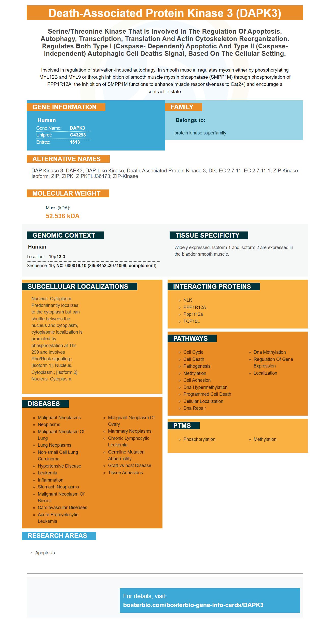
Involved in regulation of starvation-induced autophagy. In smooth muscle, regulates myosin either by phosphorylating MYL12B and MYL9 or through inhibition of smooth muscle myosin phosphatase (SMPP1M) through phosphorylation of PPP1R12A; the inhibition of SMPP1M functions to enhance muscle responsiveness to Ca(2+) and encourage a contractile state.
Human | |
---|---|
Gene Name: | DAPK3 |
Uniprot: | O43293 |
Entrez: | 1613 |
Belongs to: |
---|
protein kinase superfamily |
DAP kinase 3; DAPK3; DAP-like kinase; death-associated protein kinase 3; Dlk; EC 2.7.11; EC 2.7.11.1; ZIP kinase isoform; ZIP; ZIPK; ZIPKFLJ36473; ZIP-kinase
Mass (kDA):
52.536 kDA
Human | |
---|---|
Location: | 19p13.3 |
Sequence: | 19; NC_000019.10 (3958453..3971099, complement) |
Widely expressed. Isoform 1 and isoform 2 are expressed in the bladder smooth muscle.
Nucleus. Cytoplasm. Predominantly localizes to the cytoplasm but can shuttle between the nucleus and cytoplasm; cytoplasmic localization is promoted by phosphorylation at Thr-299 and involves Rho/Rock signaling.; [Isoform 1]: Nucleus. Cytoplasm.; [Isoform 2]: Nucleus. Cytoplasm.
DAPK3 modulates inflammation. What does this mean for hMSCs, however? How can it be used in clinical trials? This article lists some of its most common uses. Scientists can submit their results for product credits, samples, and other benefits. This article applies to all scientists world-wide. To receive product credits, you must be a biotechnology researcher.
DAPK3 regulates inflammation and is expressed in a variety cell types, including intestinal epithelial cells and innate and adaptive immuno cells as well as lymphoid tissue. Its various roles include regulation of cell death, regulation of inflammation, and a prosurvival role. It may also be a therapeutic target because it regulates inflammation. Further research is needed to determine if DAPK really acts as a modulator for inflammation.
While DAPK expression in Crohn's and ulcerative colitis has been studied, little is known regarding its role in Crohn’s. It is thought to play a key role in the transformation UC associated tumors. Although the gene is involved at both pro and antagonistic levels, it is still a promising therapeutic target. Further studies are needed to clarify its role in intestinal inflammation and homeostasis.
Death signal transfer has been linked to DAPK3's regulation by DAPK1. Other studies show that DAPK1 phosphorylates DAPK3 to promote self-phagy. DAPK1 & DAPK3 interact within a catalytic amplification loop. This may explain why they are so important for inflammation. The complex regulation of DAPK1 & DAPK2 is complex. Multiple interactions occur independently.
DAPK3 is involved in the development or hypertension in spontaneously hypertensive dogs. It mediates the inflammatory effects induced in tumor necrosis Factor. Furthermore, knockdown or inhibition of DAPK3 prevented the hypertension-inducing response in these cells. DAPK3 is also involved in the regulation autophagy and inflammation as well as the development vascular diseases including hypertension.
These results are promising but further studies are needed in order to determine the exact function of DAPK protein in various diseases. It is believed that DAPK1 (and DAPK3) play different roles in inflammatory processes. This may influence their outcome. However, DAPK3 has been implicated in the regulation a number of aspects of inflammation. It may even mediate interplays between Raf and DAPK1 along with the ERK pathway.
Another important role for DAPK includes the regulation of atherosclerosis. Although it is not clear what role it plays in atherosclerotic plaques formation, it has been shown to regulate STAT3 expression in endothelial cells. It could even play a role regulating diseases via vascular ECs. However, DAPK does not affect the regulation of STAT3 in the intestinal wall.
DAPK3 promotes proliferation of cells and prevents apoptosis for many types of cancer. Proliferation was inhibited by knockdown of DAPK3 cells. It also inhibited Wnt/bcatenin signals, which prevented NSCLC progress. Moreover, DAPK3 knockdown in cancer cells inhibits cell growth and migration. Finally, DAPK3 inhibits Wnt/b-catenin signaling in human colon cancer cell lines.
DAPK1 is a transcription factor that is implicated in multiple types of inflammation. Boster Bio DAPK3 blocks iNOS transcription and increases CD4+-T-cell proliferation, both key components of the inflammatory response. DAPK3 has anti-inflammatory properties and is known to inhibit IL-6 and SOCS1 expression.
In order to study this protein in hMSCs, lentiviral-mediated RNA interference was used to silence its expression. The cells were then cocultured in a mixture of peripheral blood mononuclear and hMSC cells for three consecutive days. After the cells had been cultured we used western blotting to determine DAPK1's expression. Other experiments included the detections IL-6, IL-10, SOCS1, and SOCS2. Flow Cytometry was used to assess the proliferation of CD4+T cells.
The best use of DAPK3 marker hMSCs is modulation in expression of ABCA7/PLOD1. These genes are involved both in neurodegeneration as well as aging. Boster antibodies are high-affinity, validated for multiple applications across Western Blotting, Immunohistochemistry, and ELISA. The first reviewers of a product receive product credits from Boster, giving them a leg up on their competitors.
Research may benefit from using DAPK3 gene expression to guide iPSCs. It is a good indication that MSCs express these genes differently. It is important to note, however, that MSC expression of most iPSC DEGs has been higher than in iPSC. This means that genes with high expression in iPSC and hMSC are enriched in lineage-relevant groups.
PMID: 9488481 by Kawai T., et al. ZIP kinase, a novel serine/threonine kinase which mediates apoptosis.
PMID: 10356987 by Murata-Hori M., et al. ZIP kinase identified as a novel myosin regulatory light chain kinase in HeLa cells.