This website uses cookies to ensure you get the best experience on our website.
- Table of Contents
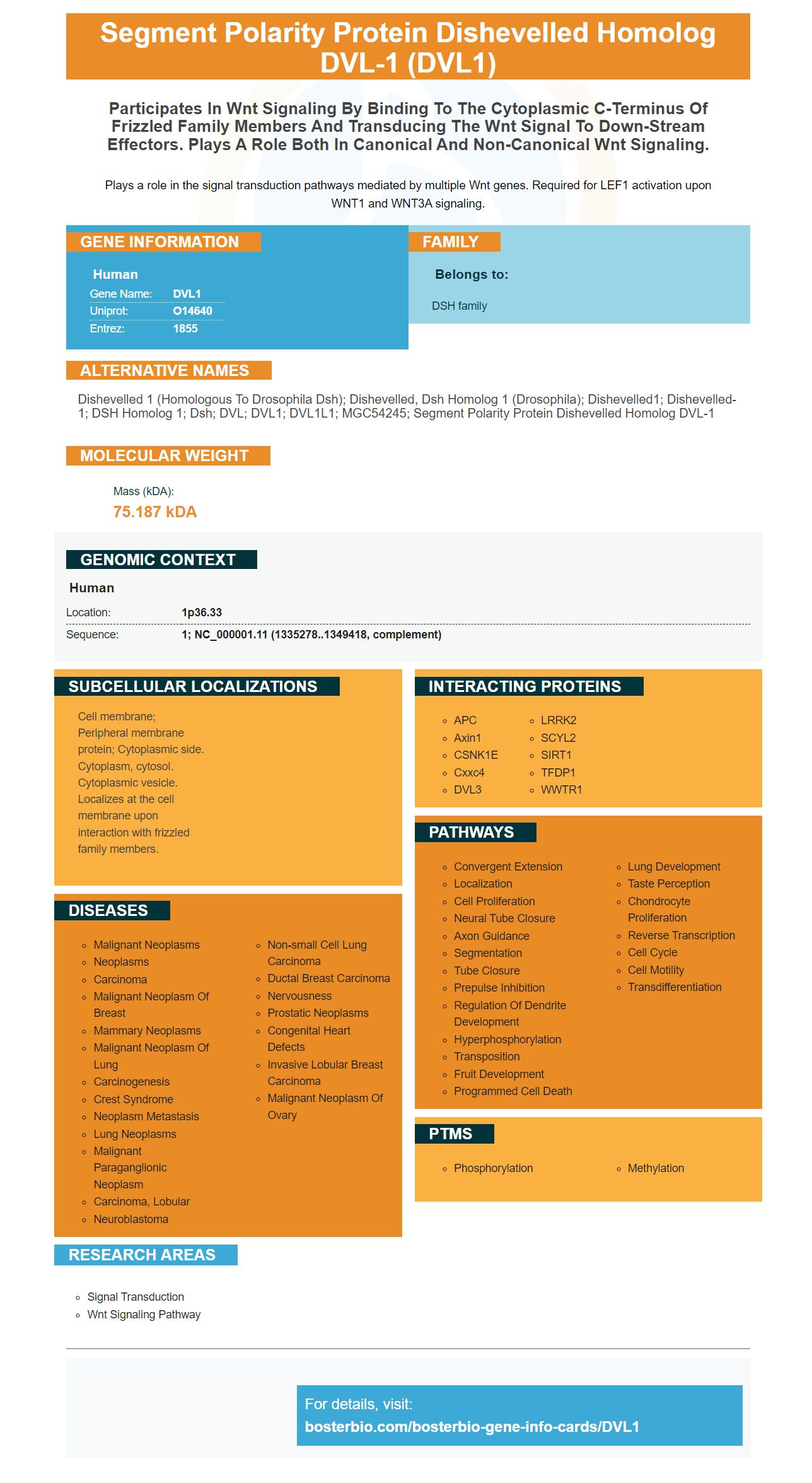
Facts about Segment polarity protein dishevelled homolog DVL-1.
Plays a role in the signal transduction pathways mediated by multiple Wnt genes. Required for LEF1 activation upon WNT1 and WNT3A signaling.
Human | |
---|---|
Gene Name: | DVL1 |
Uniprot: | O14640 |
Entrez: | 1855 |
Belongs to: |
---|
DSH family |
dishevelled 1 (homologous to Drosophila dsh); dishevelled, dsh homolog 1 (Drosophila); Dishevelled1; Dishevelled-1; DSH homolog 1; Dsh; DVL; DVL1; DVL1L1; MGC54245; segment polarity protein dishevelled homolog DVL-1
Mass (kDA):
75.187 kDA
Human | |
---|---|
Location: | 1p36.33 |
Sequence: | 1; NC_000001.11 (1335278..1349418, complement) |
Cell membrane; Peripheral membrane protein; Cytoplasmic side. Cytoplasm, cytosol. Cytoplasmic vesicle. Localizes at the cell membrane upon interaction with frizzled family members.
Antibodies to DVL1 Marker antibodies can be used for many research experiments including immunohistochemistry or Western blotting. This article will give an overview of DVL1 and its uses. This article will help you choose the right internal control protein for your research. Continue reading to learn more. Boster offers high affinity primary antibodies with a long history of being cited in the scientific community. These antibodies are not only trusted and cited by researchers but also ELISA and Immunohistochemistry approved.
The DVL1Marker can be used for the detection of cancer cells in various tissues. This marker is found in many types of cancer, including soft tissue tumors that are childhood-onset. Its high level of expression has been linked to the growth of skeletal muscle cancers. Many cancers have high DVL proteins levels. Targeting this marker may be helpful in cancer research. However, it remains to be determined which DVL1 is the most effective for cancer research.
DVL1 is a component of WNT Signalling. However, there have been several studies that have confirmed this. The regulation of myoblast proliferation by DVL3 has also been confirmed. DVL1-3 may also influence the differentiation and growth of muscle cells. If the DVL1 gene gets knocked down, then these cells will stop growing.
Another study revealed that EphB3 expression in Dvl1-/ mice is downregulated. This suggests that Dvl1 mice may have Paneth cells misplaced in their GI tract. These findings have important implications for future research and the development of new treatments. Fortunately, DVL1 gene expression remains highly conserved in other tissues. And the gene's function in regulating Wnt signaling will be uncovered.
The DVL1 indicator has been linked to microbial manipulation of gastrointestinal microbiome. These manipulations are possible to save Dvl -associated gastrointestinal problems. Nevertheless, they are not yet fully understood. Further studies will be needed to prove the benefits associated with microbial manipulation of GI microbiomes.
The DVL1 marker, an immunotherapy target, is responsible for enhancing the immune response in tumor cells. It is present in many cancers including lung, breast and prostate. These treatments are designed to target the immune system and have multiple therapeutic benefits. Researchers are currently trying to identify immunotherapeutic targets by evaluating a patient's immune response. Newer methods are available to measure these markers.
Monoclonal antibody treatments that target inhibitory immune cells, such as PD-L1, are effective for treating a variety of cancer types. Both monomodality and combination immunotherapies require better biomarkers to guide the course of therapy. The Immune Report Card is one such biomarker. It measures the expression of key immune response cycles markers and immunotherapeutic targets. It provides a comprehensive picture and analysis of the patient's immune responses.
The DVL1Marker has been shown that it can localize to genes associated with cancer and regulate their expression. The findings suggest that DVL-1 may be a key regulator of the Wnt signaling pathway. It is notable that this protein is also expressed within normal breast tissue (including the ductal Adenocarcinoma). It is important to note that although this protein is important in cancer biology, its expression is not controlled in normal tissues.
DVL-1 (a nuclear scaffold protein) acts as a transcription factors and repressor. Interestingly, DVL-1 is expressed in tumors differently in different types of cancers. It regulates the expression targets genes through its conserved amino acid sequences. Future studies will use these data to determine the role that this gene plays in tumor growth. You can also visit our website for more information about DVL-1's function.
This study also suggests that DVL1 may play an important role in TNBC-related pathology. In addition, it is important to identify unexpected gene targets in DVL-1, as this protein is implicated in directing Wnt signaling to cancer cells. This preliminary study is not conclusive, and further research is needed in order to confirm the role played by DVL in TNBC-related tumourigenesis.
Triple-negative breast cancer is linked to DVL1. Studies have shown that DVL1 regulates cellular functions and modulates the tumor's microenvironment. Moreover, DVL-1 levels correlate negatively with the tumor's immune cells. This makes it an ideal candidate to be a biomarker for this type of cancer. There are many unmet therapeutic needs.
PMID: 9192851 by Semenov M.V., et al. Human dishevelled genes constitute a DHR-containing multigene family.
PMID: 11742073 by Chen W., et al. beta-Arrestin1 modulates lymphoid enhancer factor transcriptional activity through interaction with phosphorylated dishevelled proteins.