This website uses cookies to ensure you get the best experience on our website.
- Table of Contents
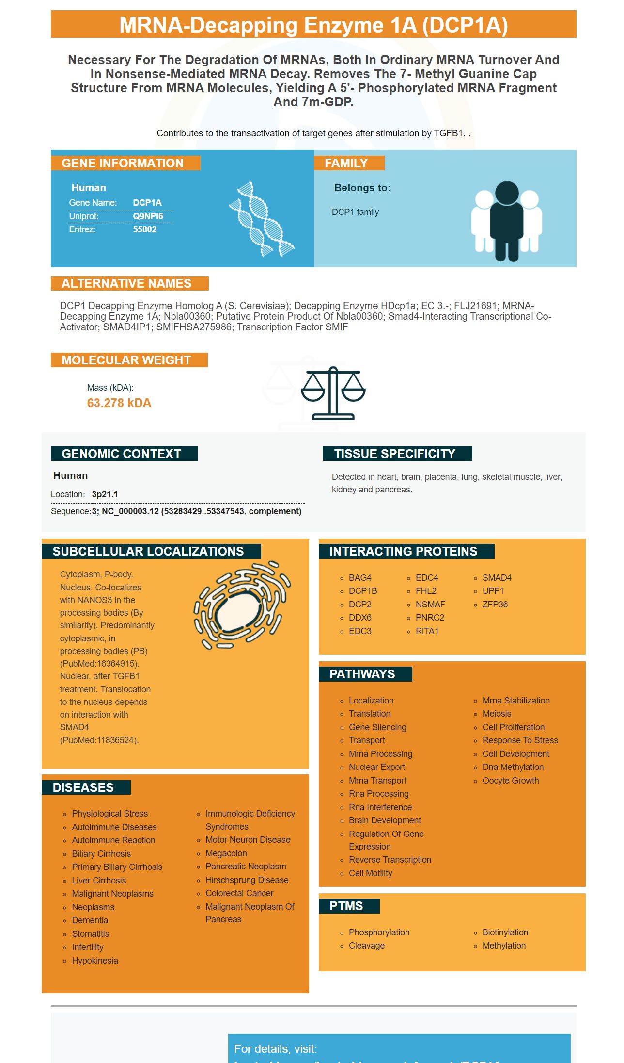
Facts about mRNA-decapping enzyme 1A.
Contributes to the transactivation of target genes after stimulation by TGFB1. .
Human | |
---|---|
Gene Name: | DCP1A |
Uniprot: | Q9NPI6 |
Entrez: | 55802 |
Belongs to: |
---|
DCP1 family |
DCP1 decapping enzyme homolog A (S. cerevisiae); decapping enzyme hDcp1a; EC 3.-; FLJ21691; mRNA-decapping enzyme 1A; Nbla00360; putative protein product of Nbla00360; Smad4-interacting transcriptional co-activator; SMAD4IP1; SMIFHSA275986; Transcription factor SMIF
Mass (kDA):
63.278 kDA
Human | |
---|---|
Location: | 3p21.1 |
Sequence: | 3; NC_000003.12 (53283429..53347543, complement) |
Detected in heart, brain, placenta, lung, skeletal muscle, liver, kidney and pancreas.
Cytoplasm, P-body. Nucleus. Co-localizes with NANOS3 in the processing bodies (By similarity). Predominantly cytoplasmic, in processing bodies (PB) (PubMed:16364915). Nuclear, after TGFB1 treatment. Translocation to the nucleus depends on interaction with SMAD4 (PubMed:11836524).
In this article, you'll find out about DCP1A, an mRNA-decapping enzyme 1a. This enzyme can be used to detect colorectal cancer. It can also be used as a highly sensitive diagnostic indicator. It could be a sign of a bad prognosis if it's high in the tissue of colorectal carcinoma. Learn more about how this marker can be utilized in your practice.
The enzyme that degrades mRNA is a crucial component of eukaryotic degrading mRNA, that acts as a regulator of gene expression. Boster Bio's 1A MRNA-decapping Protein can be used to analyze the cellular processing bodies (or PBs). Cancer is often associated with PBs. The protein decapping enzyme for mRNA is involved in the physiology and development of cancer cells.
The expression of DCP1A was determined using reverse transcription quantitative polymerase chain reaction (RT-qPCR) and the Student's t-test. The relative expression level of DCP1A was significantly higher in colorectal cancer tissue than in non-carcinoma tissues. The experiments were repeated three times to confirm the results. DCP1A was significantly higher in tumors than it was in healthy tissues.
The levels of expression of DCP1A in non-cancerous tissues was compared, and DCP1A expression was classified as either low or high. After these measurements, clinicopathological parameters were collected and the differences were analyzed using the kh2 test. Age and gender did not influence DCP1A expression. The high expression of DCP1A in CRC was associated with greater invasion and greater lymph node metastasis.
Exogenous mRNA can be delivered in the vivo. This is a major challenge. The focus of research has been on the development of more efficient delivery methods for exogenous mRNA. We don't discuss specific delivery platforms, but we will provide readers with excellent reviews. We look forward to Boster Bio's further development of its mRNA-decapping enzyme 1a marker.
Non-structural genes, subgenomic promoters and a variable Goli Index are all part of MRNA self-amplifying. This variable GOI replaces the codon sequence of viral structural proteins. Self-amplifying mRNA is stable and exhibits a broad spectrum of gene expression. Boster Bio's mRNA decapping enzyme 1a indicator is extremely antiviral.
The iCycleriQ Real Time Detection System for DCP1a is a high-performance iQ real-time PCR detection system with flexibility to suit multiple applications. The system includes the thermal cycler 96 well with an integrated optical module. This module is able to connect the sensitive filter-based optics to the 96 well plate. This system also comes with a fan-cooled 50-watt tungsten halogen lamp. The optical module has the 6 position filter wheel and the dual-mirror arrangement.
DCP1a expression is linked to poor prognosis among colorectal carcinoma patients. However, it is still not clear if DCP1a is an indicator for diagnosis or is an indicator of the prognosis of colorectal cancer. This system is a crucial element of your laboratory's testing procedure. It provides many advantages.
One of the most popular protein detection methods is immunohistochemistry. This technique utilizes antibody-antigen interactions to visualize the distribution of specific cellular components. Researchers optimize the staining and preparation of samples to create a strong signal. The results of successful experiments can be evaluated by the scientific community. The Sensitive diagnostic marker from Boster Bio can be used in a variety of fields of medicine. The company is committed to making the detection of cancer easier and more precise than ever before.
The IL6 marker from Boster Bio has excellent predictive value when utilized in conjunction with other diagnostic tests. It is highly prognostic when used in the setting of antibiotic resistance, and is recommended for use alongside other tests by healthcare professionals. Independent raters have evaluated it for accuracy and detection window. The short half-life, as well as the narrow detection window limit its use as a monitoring technique.
The PDGF-R-based IHC test is another sensitive biomarker. The Sensitive Diagnostic Marker is a biomarker used for several diseases, including cancer, inflammation, and neurology. It is extremely sensitive and can identify just one picogram. Boster Bio products for immunology can be purchased via tebu.bio. The company also offers an array of secondary antibodies, isotype controls and detection systems designed for IHC.
Colorectal cancer patients have a poorer prognosis when they have DCP1A. It can also be used as a diagnostic or prognostic marker. Recent research suggests that the DCP1A protein could play a role in the formation of P-body as well as carcinogenesis. These findings could help in the development of new treatments.
Researchers discovered a significant correlation between CXCR7 gene expression and overall survival. The correlation was stronger in patients suffering from cancers of stage IV or greater. Additionally patients with low levels CXCR7 had low overall survival. The study also showed that low levels of CXCL12 expression was associated with low differentiation, and local and lymphovascular invasion as well as advanced stages of the disease.
Recent studies have demonstrated that DCP1A plays an important role in the progression of colorectal cancer. Epigenetic changes, including suppression of the DCP1A gene, are involved in the formation and progression of colorectal cancer. Epigenetic regulation plays a crucial role in the development and progression of cancer. Therefore, identifying the DCP1A marker in colorectal tumors will allow medical professionals to identify patients who are at risk for resection.
The expression of DCP1A in colorectal cancer shows positive correlation with E-cadherin levels and metalloproteinase-9 activity, and E-cadherin. The study also indicates an immediate relationship between TGFB1 and DCP1A expression. The authors also note that DCP1A has a direct role in colorectal cancer progression.
The DCP1A gene is a crucial gene regulatory protein that is involved in mRNA degradation. DCP1a can also be used to determine cellular processing bodies that are associated with various ailments and conditions. It has also been associated with a higher incidence of melanoma, which is a type of colorectal cancer. It also plays a role in the progression of cancer.
A recent study showed that the CD44 variant of the DCP1A gene regulates oxidative stress in cells. It interacts with a subunit of the cystine/glutamate transporter, promoting cystine uptake. This is a way to increase the growth of tumors, metastasis and resistance to chemotherapeutics.
PMID: 12417715 by Lykke-Andersen J.; Identification of a human decapping complex associated with hUpf proteins in nonsense-mediated decay.
PMID: 11836524 by Bai R.-Y., et al. SMIF, a Smad4-interacting protein that functions as a co-activator in TGFbeta signalling.