This website uses cookies to ensure you get the best experience on our website.
- Table of Contents
31 Citations 16 Q&As

42 Citations 16 Q&As

26 Citations 16 Q&As
24 Citations 4 Q&As

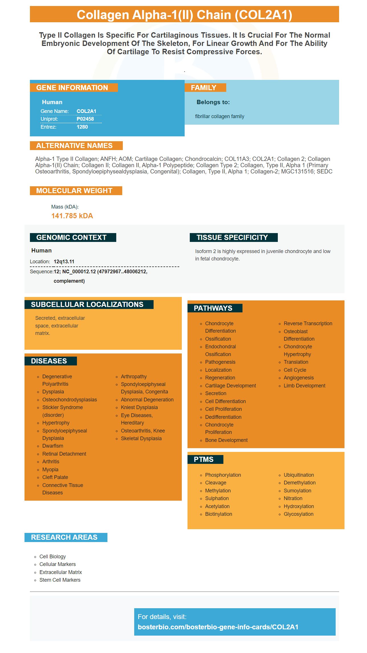
Facts about Collagen alpha-1(II) chain.

.
| Human | |
|---|---|
| Gene Name: | COL2A1 |
| Uniprot: | P02458 |
| Entrez: | 1280 |

| Belongs to: |
|---|
| fibrillar collagen family |

Alpha-1 type II collagen; ANFH; AOM; cartilage collagen; Chondrocalcin; COL11A3; COL2A1; Collagen 2; collagen alpha-1(II) chain; Collagen II; collagen II, alpha-1 polypeptide; collagen type 2; collagen, type II, alpha 1 (primary osteoarthritis, spondyloepiphysealdysplasia, congenital); collagen, type II, alpha 1; Collagen-2; MGC131516; SEDC
Mass (kDA):
141.785 kDA

| Human | |
|---|---|
| Location: | 12q13.11 |
| Sequence: | 12; NC_000012.12 (47972967..48006212, complement) |
Isoform 2 is highly expressed in juvenile chondrocyte and low in fetal chondrocyte.
Secreted, extracellular space, extracellular matrix.


What are the best uses of COL2A1, a molecular Marker for Arthritis? This article will cover these questions and more. The most effective uses for this COL2A1 marker will depend on what you are trying to measure. This article will go over Coomassie blue staining, Normalization to total protein, applications, and specificity.
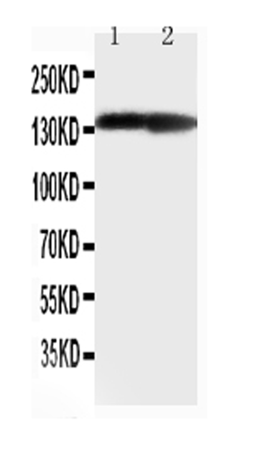
Coomassie brilliant Blue staining can be used to visualize the transfer of proteins between gels. Coomassie brilliant blue staining can be used to detect protein in concentrations up to 200 ng. It can also be used to identify imperfections in blots, such as bubbles and uneven transfers. It is important to note that Coomassie brilliant blue staining can only be used for PVDF membranes.
Coomassie brilliant is a well-known staining marker, which is cost-effective and affordable. It binds proteins non-covalently through hydrophobic and ionic interactions. It is extremely sensitive and causes the appearance of dark bands against a light blue background. Coomassie brilliant blue can also be used to detect proteins in tissues or cells.
The gel's concentration can be adjusted to improve transfer of protein. Higher concentrations require longer transfer times. A greater concentration of gels could enhance the efficiency of transfer of proteins. The primary antibody binds to the target protein. The secondary antibody binds the antibody, and then is incubated with it. This helps to detect bands of protein in western-blotting. Researchers employed this staining marker in a recent study to find protein fragments in the mouse brain.
The COL2A1 protein is a popular marker to detect collagen, a kind of cytoskeleton. We employed ACTB and GapDH as reference genes to normalize COL2A1 levels to the total protein in our study. These two genes are regarded to be stable reference genes. Additionally they have average Ct values are lower than 0.1, making them suitable as reference genes.
During chondrogenic differentiation we examined markers associated with the phenotypic differences of BM-MSCs. After 6 days of micromass-culture in an inductive medium that contained BMP2, we observed increased levels of these markers. Interestingly, all normalization methods demonstrated similar levels of expression which included single reference genes as well as a mixture of stable reference genes. We also employed a geometric average of Mon2 and Ap3d1 as a well as Fbxw2.
We utilized multiple gene panels and Proteome Discoverer software version 2.2.2 to search for the gene. We utilized the Sequest search engine to search the Swiss-Prot database Mus musculus version 2017-10-25. We selected a few genes that were expressed in C3H20T1/2 cells varieties, including GAPDH, 18S and ActB.
The COL2A1 marker is believed to be highly specific for osteoarthritis. It has the ability to identify damaged cartilage in mice models. It is also sensitive for p-SMAD1S463/465 and COL10A1. Recent studies have shown that silencing COL2A1 significantly decreased p-SMAD1S463 which is a molecule that regulates Smad1/ERK1/2.
COL2A1 regulates BMP/SMAD1 signals within chondrocytes. This receptor also inhibits SMAD1 activation and nuclear import. In the presence of COL2A1, SMAD1 expression is reduced in cartilage degenerative in humans. Osteoarthritis is likely to be caused through the degrading of COL2A1. However, despite these findings, it is not clear what function COL2A1 plays in OA progress.
The study evaluated the expression levels of 84 genes within the COL2A1 pathway. In particular we identified 26 genes that had significantly elevated expression levels in homozygous mice. We also utilized annotations of genes to determine the biological processes that are most often overrepresented. We identified "ossification" and "bone development" as the two biological processes that are most frequently overrepresented. processes. The most useful gene was "Positive Regulation of Cell Differentiation".
Multiple families were examined and found not to be associated with COL2A1. One family was not affected by severe myopia or retinal detached. These results are consistent with other genetic variations. However, despite these differences, the COL2A1 gene has been widely used to detect an inherited eye disease. It is a helpful indicator in the early detection and diagnosis of eye disease in this field.
We also discovered COL2A1 within the cochlea . We also investigated the effects of this gene on the functioning of the spiral ligament. We also observed that mice with a transgenic of the Col2a1 gene background showed abnormal type II collagen fibrils in the inner ear, which could cause an additional degeneration of the SGNs. At P150, the cochlea morphology in mice carrying Col2a1Loxl3 was normal. However, those who were knocked out of the gene had progressive hearing loss.
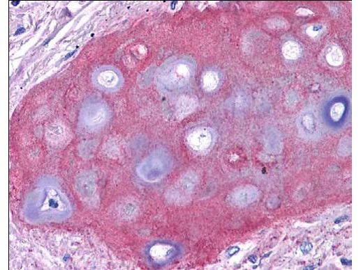
The COL2A1 marker is an important matrix protein, which is expressed predominantly in the cartilage of the articular region. This marker can be a marker of chondrogenic potential and fate, despite being a late marker of Chondrogenesis. Differences in the COL2A1 marker could impact the cell types, differentiation pathways subpopulations, as well as other functions. These findings have significant implications for chondrogenesis.
SOX9 is a transcriptional factor that is involved in the formation of cartilage and regulates the COL2A1 gene. It is also a potent activater of type II collagen, a phenotypic marker of cartilage the chondrocytes. SOX9 overexpression also increases the expression of the COL2A1 gene. This is a significant factor for osteoarthritic cartilage, since the COL2A1 gene is essential for the articular chondrocytes' differentiation. SOX9 cannot restore the osteoarthritic chondrocyte phenotype on its own.
Researchers have discovered distinct gene expression patterns in sorted and unsorted chondroprogenitor cell lines by using COL2A1-tagged chondroprogenitor cell lines. They also determined the levels of SOX9 expression and the expression of COL2A1. While the COL2A1 marker does not enhance overall gene expression, sorting can affect the number of chondrogenic cells present in the sample.
Many rare autosomal dominant conditions have been linked to the COL2A1 genetics, including type II collagenopathies. There are more than 400 mutations reported in public databases, of which 329 have been classified as pathogenic and 153 are of uncertain significance. There are three kinds of mutations: point mutations, deletions and insertions. Complex rearrangements are also possible. This mutation type determines COL2A1 inheritance pattern.
PMID: 2587267 by Su M.W., et al. Nucleotide sequence of the full length cDNA encoding for human type II procollagen.
PMID: 8948452 by Ala-Kokko L., et al. Conservation of the sizes of 53 introns and over 100 intronic sequences for the binding of common transcription factors in the human and mouse genes for type II procollagen (COL2A1).
*Showing only the more recent 20. More publications can be found for each product on its corresponding product page