This website uses cookies to ensure you get the best experience on our website.
- Table of Contents
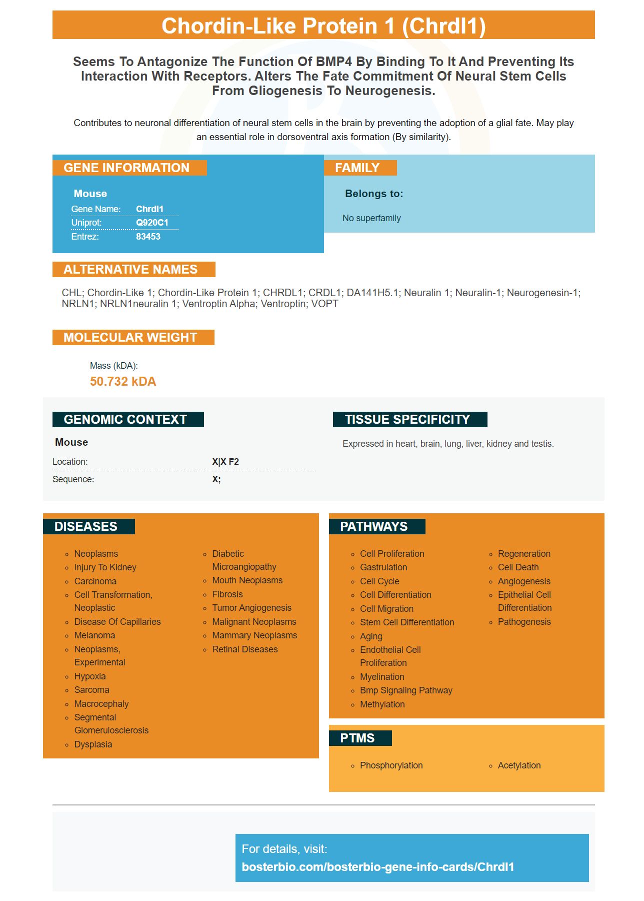
Facts about Chordin-like protein 1.
Contributes to neuronal differentiation of neural stem cells in the brain by preventing the adoption of a glial fate. May play an essential role in dorsoventral axis formation (By similarity).
Mouse | |
---|---|
Gene Name: | Chrdl1 |
Uniprot: | Q920C1 |
Entrez: | 83453 |
Belongs to: |
---|
No superfamily |
CHL; Chordin-like 1; chordin-like protein 1; CHRDL1; CRDL1; dA141H5.1; Neuralin 1; neuralin-1; neurogenesin-1; NRLN1; NRLN1neuralin 1; Ventroptin alpha; ventroptin; VOPT
Mass (kDA):
50.732 kDA
Mouse | |
---|---|
Location: | X|X F2 |
Sequence: | X; |
Expressed in heart, brain, lung, liver, kidney and testis.
The CHRDL1 marker can be used in many ways in fibrosis research. You can use it alone or in combination to predict advanced fibrosis. This article will cover the best uses of this marker as well as why it is so useful in liver fibrosis study research. Learn more about how it can be used to interpret biological samples.
The CHRDL1Marker is a protein which regulates bone remodeling and repopulation. This protein is found on the outer medulla's proximal tubules. It is not detectable in the medulla. In immunofluorescence, the expression of pSmad1/5/8 is lower than in immunohistochemical assays, but the signals it signals are more easily visible. This marker is most prominent in the S3 segment (proximal tubule), which is frequently affected with ischemic injury.
This protein binds BMP7 and antagonizes BMP7 in the presence TWSG1. CHRDL1 amplifies BMP7 signaling and is not dependent on BMP7's physical association. CHRDL1 is also an antagonist of BMP7 when TWSG1 exists, and it continues to be a BMP4 amplifier when TWSG1 exists.
In HK-2 and HEK293 cell lines, Chrdl1 was identified as a fibrosis marker. RTPCR using recombinant IgG (IgG) and recombinant CHRDL1 (mIMCD-33) was used to detect immunoprecipitated protein. DNAsedRNA (generic DNA) and genomic DNA (negative controls) were used as the positive and negative controls, respectively. In situ hybridization revealed CHRDL1 proteins were expressed on the cortex, cortex and proximal tubules.
CHRDL1 expression is actually lower after ischemia. In fact, Chrdl1 expression dropped to 5% in 24 hours after ischemia. This result was verified by pooling RNA from three animals. It was interesting to note that when BMP antagonists were tested, a reduction in Chrdl1 expression in five samples taken from injured kidneys was observed.
The CHRDL1 marker is able to detect specific cell types or genes in humans. CHRDL1 was immunoprecipitated using conditioned medium from HK-2 cells or HEK293 Cells. Recombinant protein were incubated overnight with a goat anti-CHRDL1 polyclonal antibody and Protein G sepharose beads at 4°C. The protein was then immunoprecipitated with a monoclonal antihuman antibody.
Immunostaining of P19 or MDCK cells with CHRDL1 significantly reduces BMP signaling in the developing kidney. CHRDL1 has been shown to inhibit BMP7 activity and antagonize BMP7's antagonistic properties. These findings suggest CHRDL1 may still function in adult kidneys.
The CHRDL1 marker can also be used in combination with other markers to detect BMP7. In co-immunoprecipitation experiments, CHRDL1 binds BMP7 in a dosage-responsive manner. In co-immunoprecipitation experiments, CHRDL1 and BMP7 are co-reactive in a manner that depends on the presence of TWSG1.
Chrdl1 is expressed within the medullary, proximal tubules and human renal medulla. CHRDL1 co-localizes with lotuslectin when it is stained. The antibody used to detect CHRDL1 co-localizes with lotus lectin in the proximal tubule cells.
There is increasing evidence that ceRNAs regulate gene networks. A thorough review of all known algorithms was conducted to identify the modulators. Many studies didn't know the status of the samples' protein expression or their mutation status. This study used a weighted testing to identify differentially expressed gene. It was found STED images that were modified were significantly more informative then those that were not modified.
Noninvasive tests to measure levels of procollagen type I carboxy-terminal peptide (PICP) are limited in their ability to predict disease stage and severity in patients. The serum PICP level is elevated in 50 percent of patients with advanced liver fibrosis and liver cirrhosis, but PICP levels cannot reliably detect the disease.
Although liver biopsy is the gold standard for staging liver fibrosis, this traditional diagnostic method has been challenged over the past decade by the development of noninvasive methods that evaluate scarring in the liver. Some noninvasive techniques were validated in hepatitis C cases. Despite these improvements, the diagnostic criteria for liver fibrosis remain imprecise.
These cells were sequenced using genomic sequencing. Genomic sequencing revealed that the fragment of 4.0 kbp was significantly reduced in 5 positive lines. The fragment of 3.4 kbp was smaller than expected for targeted homologous replication. Southern analysis could only identify positive clones. Further, SacI digestion revealed a fragment that is 570 bp smaller than the native locus.
Microfibrillar-associated protein 4 (MFAP4) is a collagen-binding protein with an integrin-binding motif at the N-terminal end. It plays a role in the innate immune response, and allows for free gaseous exchanging in the lung. It also associates the collagen region with surfactant proteins.
PMID: 11441185 by Sakuta H., et al. Ventroptin: a BMP-4 antagonist expressed in a double-gradient pattern in the retina.
PMID: 11118896 by Coffinier C.C., et al. Neuralin-1 is a novel chordin-related molecule expressed in the mouse neural plate.