This website uses cookies to ensure you get the best experience on our website.
- Table of Contents
1 Citations 16 Q&As
1 Citations 5 Q&As
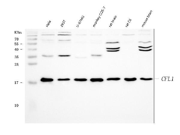
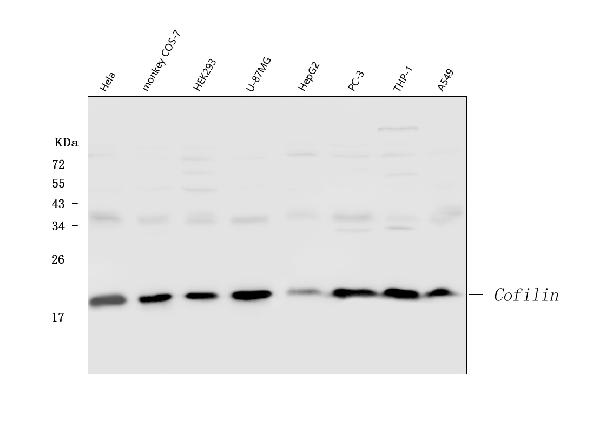
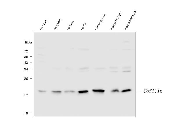
Facts about Cofilin-1.
Important for normal progress through mitosis and normal cytokinesis. Plays a role in the regulation of cell morphology and cytoskeletal organization.
Human | |
---|---|
Gene Name: | CFL1 |
Uniprot: | P23528 |
Entrez: | 1072 |
Belongs to: |
---|
actin-binding proteins ADF family |
18 kDa phosphoprotein; CFL; cofilin 1 (non-muscle); Cofilin, non-muscle isoform; cofilin-1; p18
Mass (kDA):
18.502 kDA
Human | |
---|---|
Location: | 11q13.1 |
Sequence: | 11; NC_000011.10 (65854673..65858180, complement) |
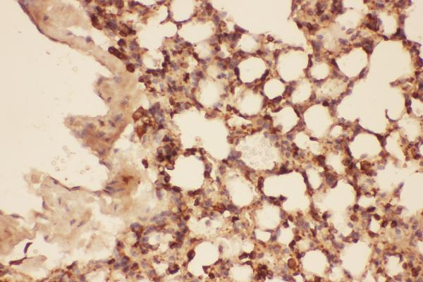
Widely distributed in various tissues.
Nucleus matrix. Cytoplasm, cytoskeleton. Cell projection, ruffle membrane; Peripheral membrane protein; Cytoplasmic side. Cell projection, lamellipodium membrane; Peripheral membrane protein; Cytoplasmic side. Cell projection, lamellipodium. Colocalizes with the actin cytoskeleton in membrane ruffles and lamellipodia. Detected at the cleavage furrow and contractile ring during cytokinesis. Almost completely in nucleus in cells exposed to heat shock or 10% dimethyl sulfoxide.
If you're in search of a biomarker to identify the CFL1 gene, you've likely come across Boster Bio's CFL1 marker. If you've been interested in purchasing this marker but aren't sure which application it's best suited for, check out some recommendations. We also discussed the cost of the Boster Bio CFL1 marker.
There are a myriad of markers, however it is crucial to pick the best one for your needs. Certain types of markers have different tip sizes than others, so you'll need to find the perfect combination of size and shape to get the job done. Double-sided markers must be stored horizontally in order to prevent the tips from drying out. For best results, make sure you check the ink type guide to determine the best one. These markers are ideal for everyday use.
Real-time RTPCR was utilized to determine the expression of CFL1 in human tissues. This was done in 30 pairs of non-cancerous and cancerous tissue. Data were examined using SigmaPlot and SPSS software. The tumor tissue samples were found to have higher levels of CFL1 than non-tumor tissue samples. These findings suggest that CFL1 expression could be a useful biomarker for tumor-related disease.
The expression of CFL1 is crucial for cancer treatment. CFL1 is crucial for cancer treatment. It causes the cell cycle stop and inhibits the proliferation of tumor cells. A mechanistic investigation revealed that CFL1 overexpression caused the growth of cancer cells and invasion to be reduced. This suppressive effect may have been due to up-regulation of let-7 microRNA. Many cancers are affected by CFL1, even though it isn't clear what function it plays in cancer.
The protein cofilin 1 is a component of the actin-depolymerization factor ADF/cofilin family. It is composed of two muscle isoforms , as well in a non-muscle counterpart. CFL1 is tiny ubiquitin proteins that has multiple functions in normal cells. It is essential for cell migration and invasion in solid tumors. Cofilin 1 is used in many areas of cancer research.
CFL1 is an actin component that controls the assembly and maintenance of dynamic stress fibres made of actin in cells. CFL1 is vital to maintain cell-cell contacts and preventing the unintentional spontaneous proliferation of Keratinocytes. Without CFL1 membrane structures could still be formed, but they are less dynamic. In spite of its importance however, it is a difficult gene to define and detect cancer.
The expression of CFL1 was associated with the presence of portal vein thrombus (PVT) and microvascular invasion (MVI). MVI is a powerful independent predictor of low survival and early recurrence. PVTT can cause distant metastasis, and increases mortality among patients with HCC. The high expression of CFL1 in cancer cells could promote invasion and migration. This research area is in the process of being explored.
While it plays a crucial role in epithelial tissue organisation however, it isn't able to compensate for the absence of CFL1 in embryonic development. Although mice lacking CFL1 are viable however, they have cornea defects in adulthood, leading to blindness and other problems. Neuronal cells can also become overdifferentiated because of the loss of conditional CFL1. The corresponding changes in the growth and migration of neuronal cells result in the lissencephaly type.
CFL1 has been identified as a marker in the progression of various cancers and cancers, including NSCLC (non-small lung cancer). This gene is involved in cell motility, cytokinesis and the process of morphogenesis. It also plays a role in the movement of cancer cells and has been linked with the esophageal squamous and vulvar cancer. The CFL1 gene is also involved in the prevention of cytoskeletal-associated defects, such as axonal development in cancer cells.
Recent research has revealed that CFL1 expression was linked with poor survival among HCC patients. It was also associated with lower OS, DFS, and other factors. It was also associated with malignant progression and poor clinical outcomes and other negative consequences. CFL1 expression is linked to lower survival rates of patients and higher rates of metastasis in clinical practice. This could help in treatment.
PMID: 2263493 by Ogawa K., et al. Coding sequence of human placenta cofilin cDNA.
PMID: 7552146 by van der Steege G., et al. A provisional transcript map of the spinal muscular atrophy (SMA) critical region.
*More publications can be found for each product on its corresponding product page