This website uses cookies to ensure you get the best experience on our website.
- Table of Contents
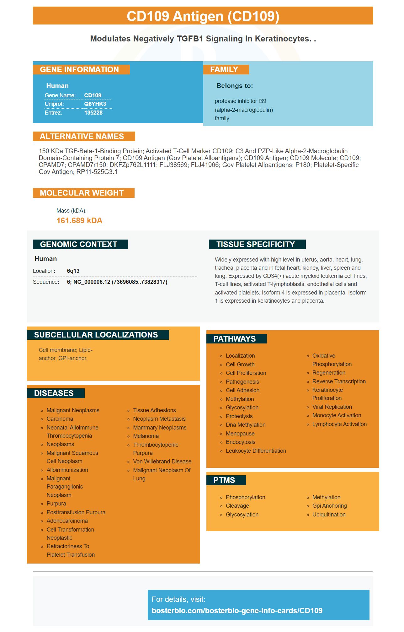
Facts about CD109 antigen.
Human | |
---|---|
Gene Name: | CD109 |
Uniprot: | Q6YHK3 |
Entrez: | 135228 |
Belongs to: |
---|
protease inhibitor I39 (alpha-2-macroglobulin) family |
150 kDa TGF-beta-1-binding protein; activated T-cell marker CD109; C3 and PZP-like alpha-2-macroglobulin domain-containing protein 7; CD109 antigen (Gov platelet alloantigens); CD109 antigen; CD109 molecule; CD109; CPAMD7; CPAMD7r150; DKFZp762L1111; FLJ38569; FLJ41966; Gov platelet alloantigens; p180; Platelet-specific Gov antigen; RP11-525G3.1
Mass (kDA):
161.689 kDA
Human | |
---|---|
Location: | 6q13 |
Sequence: | 6; NC_000006.12 (73696085..73828317) |
Widely expressed with high level in uterus, aorta, heart, lung, trachea, placenta and in fetal heart, kidney, liver, spleen and lung. Expressed by CD34(+) acute myeloid leukemia cell lines, T-cell lines, activated T-lymphoblasts, endothelial cells and activated platelets. Isoform 4 is expressed in placenta. Isoform 1 is expressed in keratinocytes and placenta.
Cell membrane; Lipid-anchor, GPI-anchor.
qRTPCR or Simple Western can be used to determine whether a sample contains the CD109 protein. Boster Bio offers product credit for scientists who perform CD109 testing on samples from specific species or applications. It is available to all scientists worldwide. Read on to learn how this product can help you. Here are the top uses of CD109 protein.
The CD109 marker, a membrane protein, has many functions in human tumors. Its positive determinants include enhancing cell migration and proliferation, sphere formation, and anchorage-independent growth. It was also shown to reverse tumorigenic effects by silencing CD109. Its role in cell growth, proliferation, and migrating is likely to be related with phosphorylation STAT3-EGFR.
The primer sequence for the CD109 marker is GGAAGGTGCTACGAAAGGTGTGTGTGTGTGTGTGTGTTACGAAAGTAG. In this study, NTC siRNA knocked down CaSki CD109(+) cells. Cell viability was also decreased. CD109 silencing also inhibited the growth gliospheres within a methylcellulose matrix.
The CD109 genes is highly expressed in human tissues. It may be useful to determine whether cells have the CD109 gene. There are many other uses for the CD109 protein. The gene can be found in the cytoplasms of many tissues and organs including the liver, pancreas, and lungs. The CD109 gene can be described as a transcription element that enables transcriptional control.
Recent research revealed a strong correlation between CD109 and MES glioblastoma type. 17 glioblastoma data files involving 2100 patients included the profile of CD109. The highest levels of CD109 mRNA were more likely to be MES or CL. This gene has been associated with many types of tumors, including gliomas, so its presence in a cancer cell sample could be useful in identifying which type of cancer it is.
Although this marker is abundant in PSCC tissues, further research is needed before this gene can be used to identify biomarkers. Further investigations into its role in this disease may facilitate earlier diagnosis and treatment. The authors gratefully acknowledge the funding of the National Natural Science Foundation of China grant 81570255 and the Shandong Taishan Scholarship for Professor Ju Liu. It has been reported CD109 may have played a role the pathogenesis of PSCC.
High levels of CD109 signaling are associated with poor prognosis. The high CD109 expression is correlated with TGF/b/Smad signalling status, tumor grade, clinical stages, and the IHC images. TGF-b/P–Smad2 and CD109 expression in OSCC are also associated with tumor grade. Moreover, high levels of CD109 are associated with highly proliferative glioblastomas.
A recent study also suggested that silencing CD109 could reduce BTSCs in gliomas. The effect was reversed by treatment with a radiotherapy drug that inhibited CD109 expression in the tumors. Further studies revealed that CD109 inhibition reduced the proliferative capabilities of glioma cellular cells. These results suggest that CD109 is a potential therapeutic target to treat gliomas.
Previous studies have identified several biomarkers to help with PSCC. There are three biomarkers for PSCC: cyclin A1, p53, Ki-67. They have not been associated with lymph node metastases or shown significant prognostic value. In fresh surgical samples, CD109 expression in PSCC cell line cells has also been confirmed. It is a promising candidate for predicting patient's prognosis.
This method allows CD109 to be detected on a subset endothelial and platelet cells. The protein also carries human platelet antengen 15 (HPA). The SNaPshot test is used to determine a patient's HPA genotype. It uses peripheral blood stem cells and RNA. The CD109 transcript expression in these cells is then determined using real-time SYBR blue PCR. The melting curve analysis is then performed.
In the SCC cell lineage, CD109 plays a crucial role as a gatekeeper. It suppresses EMT by modulating TGFB signalling pathway. It is also thought to regulate gene transcription. Additionally, this marker has been associated with tumor-related gene expression. The resulting dataset shows CD109 plays a crucial role in the progression SCC.
Higher CD109 expression in PBSCs increased the likelihood of pairing with higher cell numbers. These results were obtained using two types of PBSC. One type of donor had higher CD109 mRNA expression than another. The CD109 marker was also present in PB before the granulocyte collection. PBSC#2 had a lower CD109 level, and had fewer satellite tumors.
Cells that expressed CD109 had increased proliferation and suppressed cell death. The parental A431 cells, on the other hand, showed massive apoptosis even at full confluence. Caspase 8 expression was elevated and Ki67 expression decreased. CD109-KO cell did not show evidence of cell dying. CD109-KO mice have cells that proliferate without suffering apoptosis.
The results of the present study also showed the association between tumor progression and CD109 expression. CD109 expression was found to be higher in lower-grade tumors than in Grade III. These findings suggest that tumors with CD109 expression are associated with low-grade disease. However, more research is needed in order to confirm this association. Patients with high levels of CD109 protein could be monitored for signs of cancer progression.
Detection of CD109 in human cancer cells may be beneficial for the development of new treatments. Although the molecular functions and causes of CD109 are not known, they have been shown to be associated with tumorigenesis. Several studies have implicated CD109 in tumorigenesis. For instance, CD109 expression is associated with increased tumor size and metastasis in glioblastoma.
EMT plays an important role for CD109. It is believed to regulate transcriptional factors and facilitate cell mobility. CD109 was detected in A549 cells. Its inhibition caused a morphological change, from a circular shape to a multi-directional pattern. Moreover, CD109 knockdown reduced the expression of mesenchymal markers including N-cadherin and E-cadherin.
The detection of CD109 expression in human tumor cells offers several benefits. CD109 expression can be used to detect tumor cells and also identify cancer-related protein. The protein belongs to the complement family and the alpha-2-macroglobulin (a2M) family. Its mature form has an internal thioester bonds (ITG), which promotes covalent bonding with adjacent molecules. The CD109 human gene is located on chromosome 6. It encodes 1445 amino acid. The protein includes a N-terminal leader sequence of 21 amino acids, as well as a putative bait area containing an asymmetrical pentapeptide and a GPI signal sequence.
The expression of CD109 was determined in human PSCC tissues using immunohistochemistry as well as tissue microarrays. Immunohistochemical staining was performed on malignant squamous and normal epidermis. The protein was expressed at a high level in malignant tumor cells. CD109 can be used in PSCC as a therapeutic target and biomarker.
CD109 expression is associated with tumor stage and degree in human epithelial tumors. This suggests that it could play a role as a factor in tumor progression. CD109 expression is found in cancer patients' circulating endothelial tissues, but not normal tissues like the skin and penile. CD109 expression was also absent from normal PSCC blood vessel, suggesting that human skin-type SCCs are squamous.
CD109 is an essential EMT protein. Real-time real-timePCR was used to determine transcriptome levels for CD109 and EMT related genes in lung carcinomas. CD109 was found to be associated with high levels of expression in lung adenocarcinomas using the Pearson correlations. This could mean that CD109 plays a role in tumor mobility as well as EMT. In lung tumors, there is a strong connection between EMT (EMT) and CD109.
In the same study, TGF-b-induced EMT was also inhibited by hBM-MSCCM. The mechanism that hBMs-MSCs inhibit growth of tumor cells has been suggested as being the anti-cancer effects of hBMs-MSCs-CM and CD109. TGFb signalling inhibition in cancer cells leads to decreased stemness and migration for SSCs.
PMID: 11861284 by Lin M., et al. Cell surface antigen CD109 is a novel member of the alpha(2) macroglobulin/C3, C4, C5 family of thioester-containing proteins.
PMID: 14980714 by Solomon K.R., et al. CD109 represents a novel branch of the alpha2- macroglobulin/complement gene family.