This website uses cookies to ensure you get the best experience on our website.
- Table of Contents
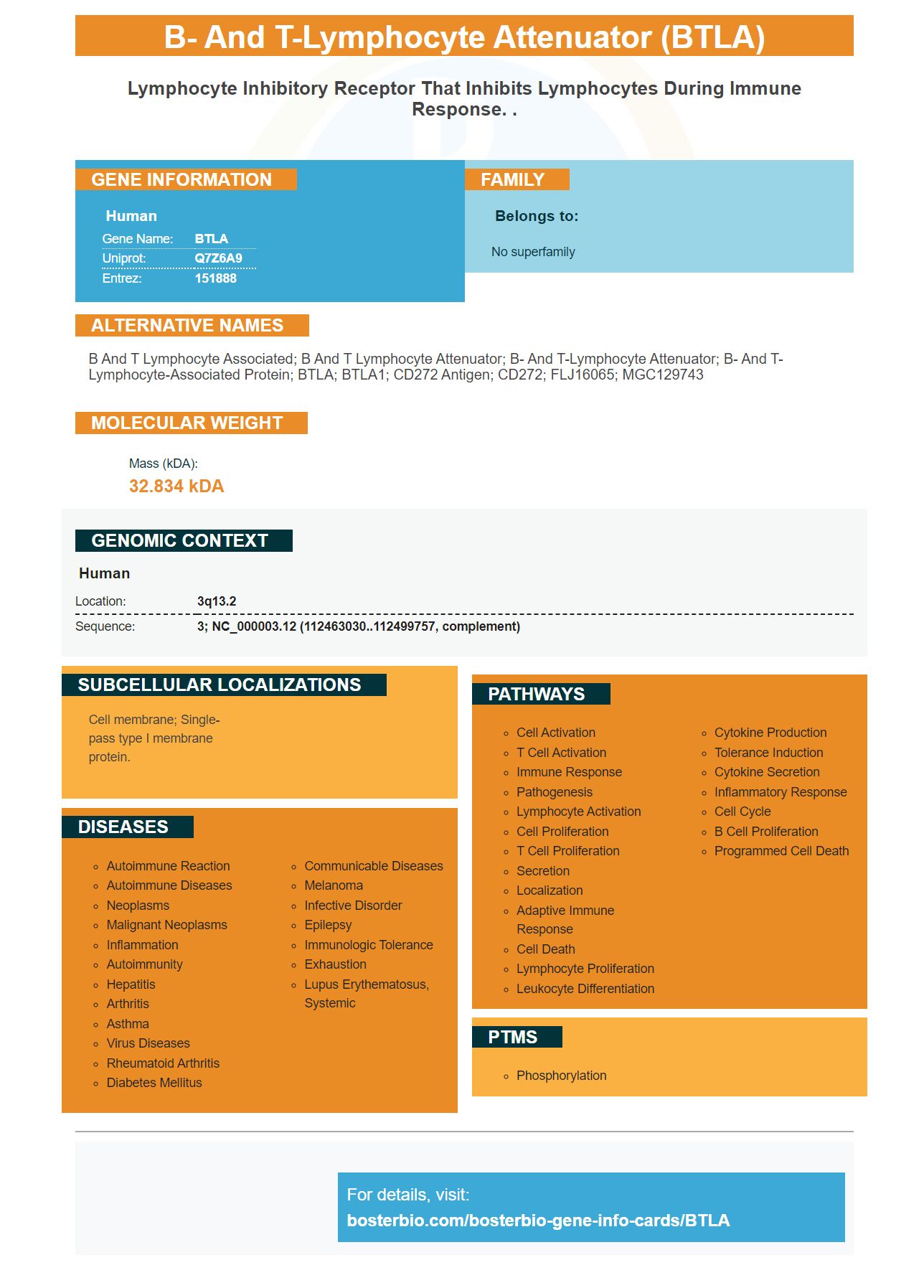


Facts about B- and T-lymphocyte attenuator.

| Human | |
|---|---|
| Gene Name: | BTLA |
| Uniprot: | Q7Z6A9 |
| Entrez: | 151888 |

| Belongs to: |
|---|
| No superfamily |

B and T lymphocyte associated; B and T lymphocyte attenuator; B- and T-lymphocyte attenuator; B- and T-lymphocyte-associated protein; BTLA; BTLA1; CD272 antigen; CD272; FLJ16065; MGC129743
Mass (kDA):
32.834 kDA

| Human | |
|---|---|
| Location: | 3q13.2 |
| Sequence: | 3; NC_000003.12 (112463030..112499757, complement) |
Cell membrane; Single-pass type I membrane protein.


BTLA is a gene with many applications and is being explored in the field of immunotherapy research. We will be discussing the function of BTLA in tumors and how it interacts with HVEM. Learn more about it. Boster Bio The Best Uses Of The BTLA Marker
The present study evaluated BTLA expression in a cohort of patients with non-Hodgkin lymphoma. The tumor samples were analyzed by immunohistochemistry with the Leica DM4000 B electron microscope. The results were analyzed using the chi-square method and Spearman's rank correlation. BTLA expression in tumor samples was calculated in terms of percent positivity (positive or negative) or as a percentage of total expression. The survival curves were calculated using Kaplan-Meier method, and Cox proportional hazards analysis was performed.
There is not much information about the role of BTLA in lung cancer. While BTLA is expressed on T cells in lung cancer its role in immune regulation is not clear. While the function of BTLA isn't well understood in the treatment of immune diseases, tumors that have higher levels of BTLA have a poor prognosis. In a recent study, Mittal et al. discovered that iNOS-induced tumors induce BTLA expression in T lymphocytes that are CD8+. These T lymphocytes show a partly dysfunctional phenotype and express BTLA as well as PD-1 on their surfaces.
The presence of BTLA was detected in 35 patients suffering from NSCLC and was associated with the amount of PD-L1 and PD-1 protein in TILs. The expression of BTLA was linked to the stage of the tumor and lymphatic invasion. A high level of BTLA was associated with a higher rate of progression. Patients who had positive BTLA expression were more PD-1-positive than those who did not have BTLA.
BTLA induction in NKT cells of type I is a characteristic of murine autochthonous malignant mammary tumors. This decreases the antitumor immune system. Upregulation of BTLA in the tumor's environment reduces the proliferation of gd T cells. Anti-BTLA monoclonal antibodies are in clinical trials to treat the disease. This antibody could hinder tumor growth by encouraging IFN-g secretion by the circulating CD4+ and CD8+ T cells.
Although BTLA has been demonstrated in studies to boost survival rates for CRC patients, further research is needed to determine the intricate mechanism behind BTLA's action in the CRC. However, BTLA may also impact the immune response of patients suffering from different types of cancers. This study provides valuable insights into BTLA and treatment for cancer.
Despite these developments, a number of studies in children with B cell precursor ALL have revealed a worse outcome. Clinical trials aren't approved for anti-PD-1 and CTLA-4 inhibitors. The effectiveness of these strategies depends on the complexity of BTLA signaling. It is possible to develop a new approach for cancer treatment. It is essential to comprehend the role played by BTLA in the development of cancer.
BTLA is an inhibitory receptor that shares similarities with CTLA-4 as well as PD-1, but differs in its function and expression. It is widely expressed and is involved in the homeostasis process of human cells. High levels of BTLA expression in tumors could be used to create new immunotherapies. It has also been proved to be effective in the treatment of lung cancer. Although more research is needed, the potential to target BTLA in tumors cannot be ruled out.
In recent studies, we have discovered that BTLA alters the activation of T cells. The autocrine IL-2 production by CD8+ T cells is essential for the secondary growth of memory cells. BTLA is well-known to regulate IL-2. We also found that BTLA interacts with HVEM, a marker for activated T cells. We also discovered a new function for LIGHT in activating CD4+ T cells.
Crosslinking to TCR is essential for BTLA and HVEM interaction. The anti-BTLA Ab inhibited T cell activation and proliferation in vitro when coimmobilized with anti-CD3. HVEM fusion protein inhibited the activation of T cells by interfering with cellular HVEM and LIGHT. However, anti-BTLA Ab did not interfere with T cell activation, however, anti-BTLA Ab prevented the binding of HVEM to BTLA.
Furthermore, BTLA affects several other pathways. It inhibits pSrc's phosphorylation S416 and the MAPK Kinase pathway. In addition, BTLA has an inhibitory effect on the p90RSK pathway. This suggests that BTLA hinders positive signaling pathways. We conclude that BTLA blocks activation of T cells by targeting the ITIM motif.
BTLA expression on Melanoma patients' PBMCs was high, and the corresponding percentages of T cells expressing BTLA were high. HVEM-expressing SKMEL37 cell did not cause cytotoxicity. Therefore, BTLA inhibits the proliferation of CD8+ T cells however it does not alter levels of TNF-a secretion.
Additionally, BTLA is necessary for IFN-g production by clones. It is required for functional inhibition of tumor-specific CD8+ T cells through BTLA-HVEM interactions. These results suggest that BTLA-expressing CD8+ cells have an essential role in the field of immunotherapy for cancer. Therefore, BTLA-expressing tumor-infiltrating lymphocytes are crucial to the suppression of cancerous cells.
In the present study, we used Melan-AMART-1-specific CD8+ T cells. The CD8+ T cells expressed BTLA in high amounts following vaccination without CpG. However, BTLA expression did not correlate with CpG vaccination frequency for EM/EMRA-specific T cells. Therefore, this study suggests that BTLA does not have an effect that is bystander to CpG vaccinations. However, despite the absence of a major effect on T cell activation by BTLA, vaccinations induced a high level of Melan-AMART-1-specific CD8+ T cells.
Expression of T-cell receptors are crucial for the production of co-stimulatory molecules such as ICOS or PD-1. The number of copies of these molecules can also affect the T-cell's function as an effector. For example, high expression of ICOS is associated with the production of IL-10 and IL-10, while lower levels of expression were linked with the most predominant production of Th2-type cytokines.
Transient transfection of AD-293 cells by a pRK5 vector was carried out in triplicate in 96-well plates. As a control, a blank pRK5 vector was used. After transfection, the cells were washed in PBS and then resuspended into human cell buffer. Alternately, the cells were treated with a stable human 3T3 BTLA cell line by retroviral infection with the MSCV-hBTLA construct.
The interplay between BTLA and HVEM in boster bio is vital for the immune system's functions. Both molecules are involved in the regulation of HIV-specific CD8+ T cells. Infected cells produce more HVEM than uninfected cells. The combination of antibody therapies that target both molecules increased the production of cytotoxic substances by T cells that effector CD8+.
The HVEM/BTLA complex is the largest and most important member of the TNF superfamily acting as a switch between co-inhibitory and stimulatory signaling in T cells. It interacts with antigen BTLA and the molecule CD160 to regulate co-stimulatory as well as inhibiting signaling pathways. BTLA has a binding affinity to HVEM and decreases T cell activation growth, proliferation, and differentiation. It also affects the synapses that cytotoxicly connect CTLs and T cells, which allows malignant cells to escape the antitumor response.
The BTLA/HVEM pathway plays a vital role in the adaptive immune system when infected with intracellular bacteria. BTLAand HVEM-deficient mouse had lower antigen-specific CD8+ T cells than mice who had normal BTLA or HVEM levels. In addition, mice treated using specific blockade BTLA/HVEM interactions had smaller OVA-specific CD8+ cells numbers than WT mice.
The LIGHT-HVEM interplay is known to participate in the pro-inflammatory and costimulation process however, the binding of HVEM with BTLA has anti-inflammatory properties. HVEM-deficient mice are more susceptible to autoimmune myelitis. Similar to a mouse model, the loss BTLA causes lung and cardiac allograft rejection.
The BTLA/HVEM fusion complex has been found to block naive t cell growth and stimulate Treg cell proliferation. Additionally, the HVEM HVEM complex is stabilized by soluble LIGHT and membrane-bound light, which disrupts the HVEM-BTLA complex. The proper functioning of the immune system is dependent on inhibition of the LIGHT-BTLA combo.
Infection-related illnesses can be avoided by taking inhibitors that hinder the interaction between BTLA and HVEM. In a mouse model of colitis the interaction between HVEM and BTLA can help prevent the development of a severe disease by blocking the destructive inflammatory response. The development of severe diseases can also be prevented through the transfer of CD4+ T cells.
The inhibition function of HVEM is essential to the proper functioning of the effector CD8+ T cells during retroviral infection. This is because HVEM blocks BTLA and CD160 interaction, which boosts the production cytotoxic molecules and the elimination of FV-infected cells. Anti-PD-L1 antibodies, which inhibit the interaction between BTLA and B cells, can also enhance HVEM-blocking antibody effects.
Recent research has shown that HVEM and BTLA regulate the survival rate of the CD8+ T cell. The immune response's various phases are affected by the interaction between HVEM and BTLA. It has been suggested that the reduced memory CD8 cell count is related to lower levels of BTLA-HVEM. The reason for the decrease in the number of memory T cells could differ from the one suggested.
PMID: 12796776 by Watanabe N., et al. BTLA is a lymphocyte inhibitory receptor with similarities to CTLA-4 and PD-1.
PMID: 14652006 by Gavrieli M., et al. Characterization of phosphotyrosine binding motifs in the cytoplasmic domain of B and T lymphocyte attenuator required for association with protein tyrosine phosphatases SHP-1 and SHP-2.


