This website uses cookies to ensure you get the best experience on our website.
- Table of Contents
1 Citations 4 Q&As
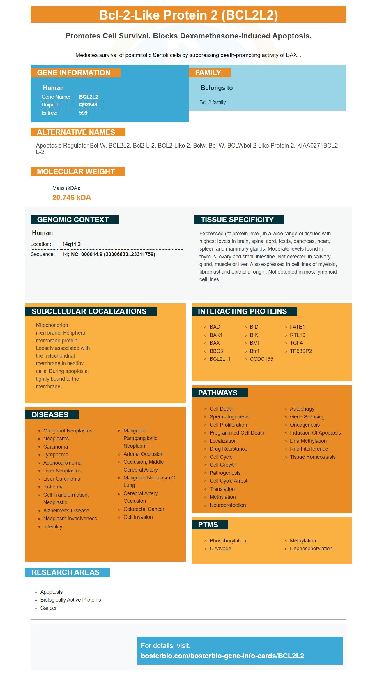
Facts about Bcl-2-like protein 2.

Mediates survival of postmitotic Sertoli cells by suppressing death-promoting activity of BAX. .
| Human | |
|---|---|
| Gene Name: | BCL2L2 |
| Uniprot: | Q92843 |
| Entrez: | 599 |

| Belongs to: |
|---|
| Bcl-2 family |

Apoptosis regulator Bcl-W; BCL2L2; Bcl2-L-2; BCL2-like 2; Bclw; Bcl-w; BCLWbcl-2-like protein 2; KIAA0271BCL2-L-2
Mass (kDA):
20.746 kDA

| Human | |
|---|---|
| Location: | 14q11.2 |
| Sequence: | 14; NC_000014.9 (23306833..23311759) |
Expressed (at protein level) in a wide range of tissues with highest levels in brain, spinal cord, testis, pancreas, heart, spleen and mammary glands. Moderate levels found in thymus, ovary and small intestine. Not detected in salivary gland, muscle or liver. Also expressed in cell lines of myeloid, fibroblast and epithelial origin. Not detected in most lymphoid cell lines.
Mitochondrion membrane; Peripheral membrane protein. Loosely associated with the mitochondrial membrane in healthy cells. During apoptosis, tightly bound to the membrane.


Boster scientists have the ability to submit their research for species, applications, or special samples. In exchange, they will receive product credits for their research. This offer is applicable to scientists all over the world. Continue reading to learn more. Below are some of the benefits of Boster Bio.
The BCL2L2 code codes for a member from the bcl-2 gene family. The gene is located in human chromosomes 14q11.2 and q12. BCL2 contains 193 amino acids. It can be found in many tissues including the colon, salivary gland, and brain. It is an anti-apoptotic regulator. It also plays a key role in cell survival when cytotoxic conditions are present.
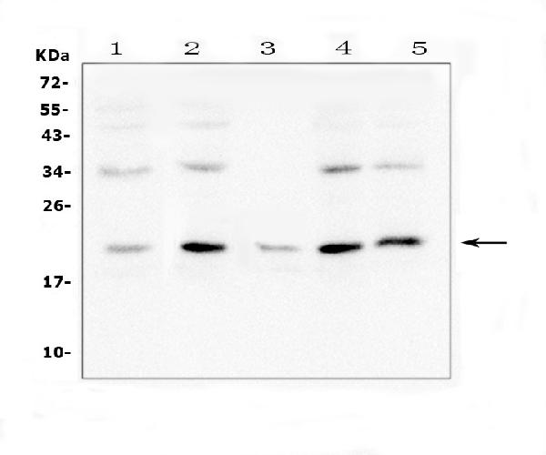
The PA1426-1 anti–BCL2L2 anti-BCL2L2 antibodies was developed and validated in a study that used human rectal cancer tissue. It was used as a Western blot analyzer and to transfer protein onto a Nitrocellulose membrane. It was used to detect the proteins using a Beyotime kit for chemiluminescence. This kit is based off boster's clone AB282.
The BCL2L2 marker is a promising biomarker for the detection of apoptotic cells in human cancer. It has been shown that megakaryocyte apoptosis is decreased when BCL2L2 is overexpressed. It also increases the number of CD41a+ LLG cells and promotes proplatelet formation. A study of 154 healthy donors found a positive correlation between platelet BCL2L22 mRNA levels and platelet number. This association shows that the BCL2L2 protein promotes platelet formation.
Gene expression studies are not perfect and cannot be compared to protein expression data. The BCL2L2 indicator is a useful biomarker that can predict the likelihood of recurrence after treatment with Tamoxifen for node negative breast cancer. BCL-2 expression was assessed using reverse transcription-PCR (from fixed tumor material). In 2007, the Journal of Clinical Oncology published these results.
The largest study that involved BCL-2 in breast carcinoma is the Nottingham series. The UBC series was four clinical trials that were completed between 1970 and 1990. It included a smaller number of patients at high risk. Bcl-2 plays a significant role for prognosis, especially in the first five year after diagnosis. As an adjunct to NPI, the BCL-2 marker may have significant potential. However, prospective research on this new cancer biomarker remains necessary.
Bcl-2 expression can be found in all NPI populations, but there is a difference between PPG and MPG. Patients with high levels Bcl-2 expression were more likely than patients with low levels to survive in the latter. This biomarker is useful in prognosticating cancer patients, but not enough to identify prognostic subgroups.
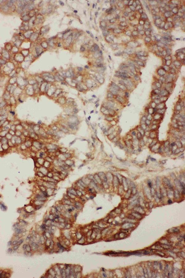
In histopathology, the BCL2L2 marker is often used to identify tumors. The marker can be used for many purposes. The BCL-2 expression in a tumor does not correlate with tumor stage, gender, or age. Bcl-2 levels and cell differentiation are not associated with Bcl-2. This study will prove its usefulness in histopathology.
A tumor may have an increased expression of BCL-w as a result of abnormal activation or signaling pathways. This protein may play a role in tumor growth and development by co-activating with oncogenes. A high level of BCL-w is a prognostic factor in cancer, although there are currently no drugs that specifically target the protein. Its expression can be used to diagnose and treat many diseases, including lung cancer and hematologic malignancies.
Although there are many potential applications of BCL2L2, few studies have demonstrated its involvement in tumor growth or aging. The gene is expressed in a wide range of cells and is associated with an increased chance of developing neurodegenerative disorders. BCL–w is also believed to promote the survival and growth of B lymphocytes. In the same way, BCL-w expression has been associated to higher levels of SOX-2, NANOG and octamerbinding transcription factor 4.
The gene can be found in many solid tissues. In tumors, BCL-w expression is associated with tumor growth in TNM stage III and poor patient survival. The protein is also associated with the presence of TNM stage III tumors and lymph nodes. This study confirmed that BCL2L2 was necessary for normal spermatogenesis. The BCL2L2 gene is found in most tumors and is highly expressed in colorectal and breast cancer cells.
BCL-2 expression was found in approximately half the NSCLCs of cancer patients. Although this is a modest number, the study reveals an important role for Bcl-2 in the treatment of NSCLC. The presence of BCL-2 in tumors may predict prognosis. In the same way, it can be used to help detect tumor growth in patients with colorectal cancer.
Molecular biology with BCL2L2, also called Bcl-w is a family protein that increases cell survival and tumorigenicity. It is expressed in many types of cancer and facilitates the production of ECM-degrading proteinases. Moreover, BCL2L2 promotes nuclear translocation of b-catenin, which increases the aggressiveness of glioblastoma (GBM) cells.
In cells, BCL-2 is expressed in response to a variety of stimuli, including cell death. BCL-2's expression levels are a key indicator of its cellular function. These levels vary from one tissue to the next. The haematopoetics system can best explain the differences in BCL-2 genes expression patterns among cells. Bclx, Mcl1, and A1 are expressed in the B lymphocyte developmental stage. Mcl1 is higher at the intermediate stage of development as the BCL-2 gene expression rises.
Quantitative real time PCR (QRTPCR) and immunoblotting were used to measure expression of BCL-2 family members. In general, BCL2L2 was downregulated in all cancer cell lines except MDA-MB-231; however, it was highly expressed in A549 and U251 cells. Additionally, the expression of miR-229b and BCL2L2 markers were ininversely related.
Solid cancers are most likely to have an amplification of MCL1 and BCL2L2 members. MCL1 and BCL2L2 are also frequently mutated in gliomas and other cancers. These gene copy number variations are a major factor in the association between these genes with BCL-2 proteins. This pattern is strongly associated to lymphomas or gliomas.
While BCL2 has been around since the 1970s and its structure is still being elucidated, it is well-known. This has led to the development of novel drugs that target the BCL-2 family. BH3 micromimetics, such as those used to treat blood cancer, have shown great efficacy. In many types of cancer, deregulations of BCL-2 proteins have been shown to be involved. By targeting pro-survival BCL-2 proteins, cancer cells may be resistant to existing treatments.
Interestingly, a mouse model failed to detect the tumour suppressive effect of Bok despite being homologous with BAK and BX. It also failed to identify the role of BOK in apoptosis. This model is sensitive and sensitive to changes in apoptosis. It may also depend on the timing for the second hit. Additionally, liver carcinogenesis is prevented by the loss of BOK in hepatocytes. However, the death of hepatocytes may promote proliferation in adjacent tissue, which is associated with mutagenesis.
PMID: 8761287 by Gibson L., et al. Bcl-w, a novel member of the Bcl-2 family, promotes cell survival.
PMID: 11423909 by O'Reilly L.A., et al. Tissue expression and subcellular localization of the pro-survival molecule Bcl-w.
*More publications can be found for each product on its corresponding product page


