This website uses cookies to ensure you get the best experience on our website.
- Table of Contents


1 Citations 1 Q&As

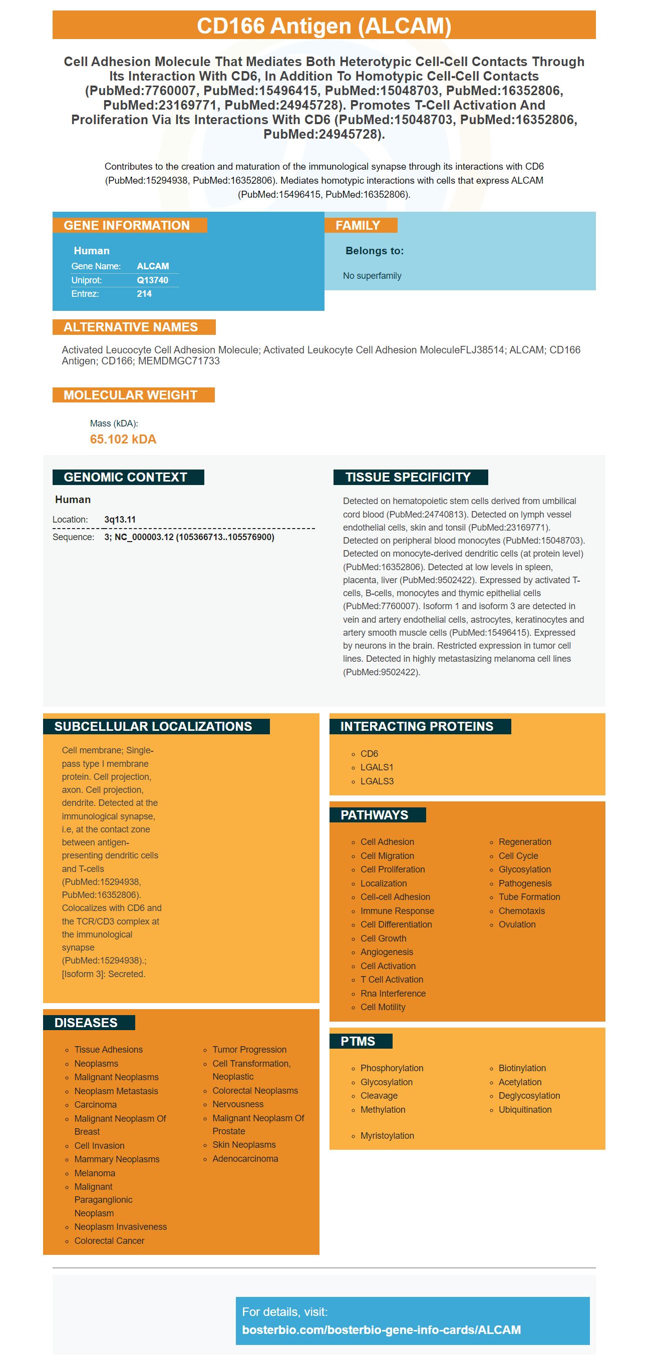
Facts about CD166 antigen.

Contributes to the creation and maturation of the immunological synapse through its interactions with CD6 (PubMed:15294938, PubMed:16352806). Mediates homotypic interactions with cells that express ALCAM (PubMed:15496415, PubMed:16352806).
| Human | |
|---|---|
| Gene Name: | ALCAM |
| Uniprot: | Q13740 |
| Entrez: | 214 |

| Belongs to: |
|---|
| No superfamily |

activated leucocyte cell adhesion molecule; activated leukocyte cell adhesion moleculeFLJ38514; ALCAM; CD166 antigen; CD166; MEMDMGC71733
Mass (kDA):
65.102 kDA

| Human | |
|---|---|
| Location: | 3q13.11 |
| Sequence: | 3; NC_000003.12 (105366713..105576900) |
Detected on hematopoietic stem cells derived from umbilical cord blood (PubMed:24740813). Detected on lymph vessel endothelial cells, skin and tonsil (PubMed:23169771). Detected on peripheral blood monocytes (PubMed:15048703). Detected on monocyte-derived dendritic cells (at protein level) (PubMed:16352806). Detected at low levels in spleen, placenta, liver (PubMed:9502422). Expressed by activated T-cells, B-cells, monocytes and thymic epithelial cells (PubMed:7760007). Isoform 1 and isoform 3 are detected in vein and artery endothelial cells, astrocytes, keratinocytes and artery smooth muscle cells (PubMed:15496415). Expressed by neurons in the brain. Restricted expression in tumor cell lines. Detected in highly metastasizing melanoma cell lines (PubMed:9502422).
Cell membrane; Single-pass type I membrane protein. Cell projection, axon. Cell projection, dendrite. Detected at the immunological synapse, i.e, at the contact zone between antigen-presenting dendritic cells and T-cells (PubMed:15294938, PubMed:16352806). Colocalizes with CD6 and the TCR/CD3 complex at the immunological synapse (PubMed:15294938).; [Isoform 3]: Secreted.


The ALCAM marker offers many benefits. It is available to scientists for species and specific studies of samples. It also applies to all scientists across the globe. The ALCAM marker can help scientists to study the expression patterns of certain cells in different tissues. It is used by Boster scientists to report their findings. These results could be used in various applications such as determining effectiveness of an immunotherapy treatment.
Researchers have discovered that the ALCAM Marker exerts an impact positive on movement of T cells and their proliferative capacity. The protein is expressed by dendritic cells of allergic asthma patients. The higher expression in biofluids from people suffering from allergic asthma and healthy controls suggests that it could be a therapeutic target. This gene is expressed on a greater proportion of T cells of patients suffering from asthma and healthy controls.
A crucial diagnostic tool is the presence of ALCAM-related markers in cancer cells. The ALCAM gene is a common target for many cancer treatments, including chemotherapy. ALCAM has been demonstrated to be sensitive to many types of cancer. Although it's not associated with survival however, it has been proven that it could help patients with advanced cancer. Researchers recently examined the survival rates of 69 patients who received chemotherapy and Tamoxifen.
Researchers also discovered that the ALCAM Marker is a positive predictor of survival rates after CRSwNP surgery. It has also been shown to be effective in separating between CRSwNP endotypes. It is crucial to remember that clinical trials results aren't conclusive. There has not been any evidence supporting the use of ALCAM to predict postoperative recurrence rates.
Patients with breast cancer can use the ALCAM Marker as a diagnostic tool. Researchers have studied more than 1,700 breast cancers for its expression. ALCAM expression was linked to rapid cell proliferation and lower survival rates in cancer cells. There are many benefits to using the ALCAM Marker in clinical trials. The results will have implications for the future of cancer research. This new marker could aid in the detection and treatment of breast cancer earlier.
ALCAM remains controversial despite its predictive value in diagnosing cancer patients. ALCAM expression has been shown to be linked with a variety of kinds of cancer. However it isn't associated with patient survival age, histology, or histology. However some studies have proven that the expression of ALCAM could indicate cancers in other tissues. The ALCAM Marker could be a new treatment option. ALCAM offers many advantages and can help with cancer treatment.
In the vivo modulating agents that are CD3 and CD6-mediated T cell activation are designed to block or induce T-cell proliferation, and vice versa. While CD3 and CD6 play similar roles but each cell type responds differently to costimulatory signals. Additionally the mAb UMCD6 inhibits the growth of T cells that are cloned. In a mouse model the mAb-induced expression of CD6 is inhibited both late and early T-cell activation responses.
The therapeutic effects that ALCAM-CD6 interaction result in the proliferation of T-cells are also very strong. These interactions are strong and long-lasting, indicating the long-term effects of CD6 on the proliferation of T-cells. Therefore, future studies will be focused on this interaction. Therefore, a new class of drugs that target CD6-mediated T cells activation is in the works.
In addition, the anti-CD3-mediated inhibition CD6 Treg cells could not result in a significant increase in the production of autoantibodies. In addition CD6-/ mice had lower levels of autoantibodies circulating. In in vivo modulators for CD6-mediated T cells' activation might be useful in increasing the circulating levels of CD8 cells and thereby improving immune responses.
Furthermore CD6-/ mice exhibit low lymphoproliferative responses to allogeneic stimulation and have a tendency to exacerbate autoimmune events. They also have elevated levels of anti-dsDNA antibodies as well as anti-nucleosome proteins, which are key indicators of Lupus-like diseases. Further evidence supports these findings in the CD6-/mice study.
Pre-treated mature DCs with 1mg/mL SEB resulted in an increase in the number of DC-T cells that contact each other. After 15 minutes at 37°C 20 percent of DC-T cell conjugates were formed. Pretreatment with SEB increased this number to 40%, suggesting that SEB enhances the formation of DC T-cell conjugates.
CTLA-4-Ig inhibits co-stimulation by blocking the interaction between CD28 and ligand. CTLA-4-Ig also inhibits both interactions. CTLA-4-Ig blocks ligands and binders in anti-CD mAb/SCFV. Both inhibit co-stimulation, but CTLA-4-Ig hinders both interactions. Both mAbs for CTLA-4Ig as well as anti-CD28 bind with Treg cells to remove them.
Anti-CD166 ALCAM Monoclonally Purified from Human Cells is available from many commercial sources. This monoclonal antibody binds to the mouse, human and rats versions of the protein. The manufacturer offers information about the antibody's immunogen, and its specificities and uses. For more information about purchasing anti-CD166 antibodies, visit the website of the vendor.
The CD166 antigen, a transmembrane glycoprotein of type I 100-105 kD, is part of the immunoglobulin family. The gene that encodes ALCAM maps to human chromosome 3q13.1-q13.2. This molecule is also referred as cluster of differentiation 166, MEMD, and SC-1/DM-GRASP in chickens. It is found in human tissues like skin and brain, melanoma, and other organs. It also serves as a barrier against autophagy and the process of apoptosis.
An Anti-CD166 ALCAM Monoclonally Purified Antibody made by Boster Bio is an excellent option for studies using this protein as an immunoassay test control. Boster has tested its antibody using various methods before releasing them. The antibodies have been thoroughly tested to make sure that they will react with the target of the cell. They are also safe to use in diagnostic tests, allowing researchers to determine whether a certain protein is present.
Boster Bio's human ALCAM/ALCAM monoclonal antibody is made to be used in conjunction in conjunction with ELISA technology. The monoclonal antibody has been precoated onto 96-well plates to maximize sensitivity and specificity. After washing the plates with PBS then a biotinylated detection polyclonal antibody from goat was added. After washing with PBS the conjugates that were not bound were removed. TMB was added following the introduction of the monoclonal antibody to detect. This kit demonstrated the HRP and TMB enzyme reaction between the human ALCAM and the standard.
The ALCAM marker is expressed in a variety of early developmental stages including the cartilage of the limbs that develop in the fetus. This marker could be used to create MSCs that are multipotent and possess differentiated specifically for the lineage. The function of ALCAM is not clear, however it has recently been linked to the development of bone in the beginning. This research is supported by the Ministry of Education.
Freshly isolated LMCs were separated to study the expression of ALCAM in the initial stages of development. This allowed us to determine whether cells that express ALCAM are ALCAM+/ALCAM-negative. Cells expressing ALCAM were examined on days 0.5, three, and five of the process of culture. This analysis was carried out using various Fc-chimeric proteins. Flow cytometric analysis was employed to calculate the percentage of ALCAM+ cells.
ALCAM was not the only marker expressed by the cell line. It also contained PDGFR Endoglin, ICAM-1 and PDGFR. ALCAMhigh cells also expressed the integrins b1 and a5 as well as the av glin. However, they did not express CD34. The cells that expressed ALCAM in the beginning of development were homogeneous in appearance and showed the expression of various cell surface markers.
ALCAM is found in the central nervous systems by microvascular endothelium. It is found in the midbrain region of the brain as well as other caudal regions. We were able identify many cell types using the anti-ALCAM antibody. The anti-ALCAM antibody identified numerous different types of cells, including eGFP+ and eGFP-positive cells.
Numerous CAMs were located in co-localization with endothelial cells and VLA-4 at day 5. VLA-4 and ICAM-1 were expressed at the same levels at both times. However, VCAM-1 markers and ALCAM markers were significantly lower. These results . After that the interactions between ALCAM and VLA-4 may be diagnostic and therapeutic targets for brain metastasis. The study concludes that ALCAM is a crucial factor in metastasis seeding.
The MACS targeting ALCAM and VLA-4 identified the presence of both proteins on LMX1A+ cells in the zebrafish embryo. The corresponding populations of mDA/GFP cells were then divided into TPBG+, double-positive, and triple-positive cells. MACS targeted the three markers produced a high proportion of cells positive (93.7 percent, TPBG-positive cell and CD4-positive cells).
PMID: 7760007 by Bowen M.A., et al. Cloning, mapping, and characterization of activated leukocyte-cell adhesion molecule (ALCAM), a CD6 ligand.
PMID: 15496415 by Ikeda K., et al. Molecular isolation and characterization of a soluble isoform of activated leukocyte cell adhesion molecule that modulates endothelial cell function.
*More publications can be found for each product on its corresponding product page