This website uses cookies to ensure you get the best experience on our website.
- Table of Contents
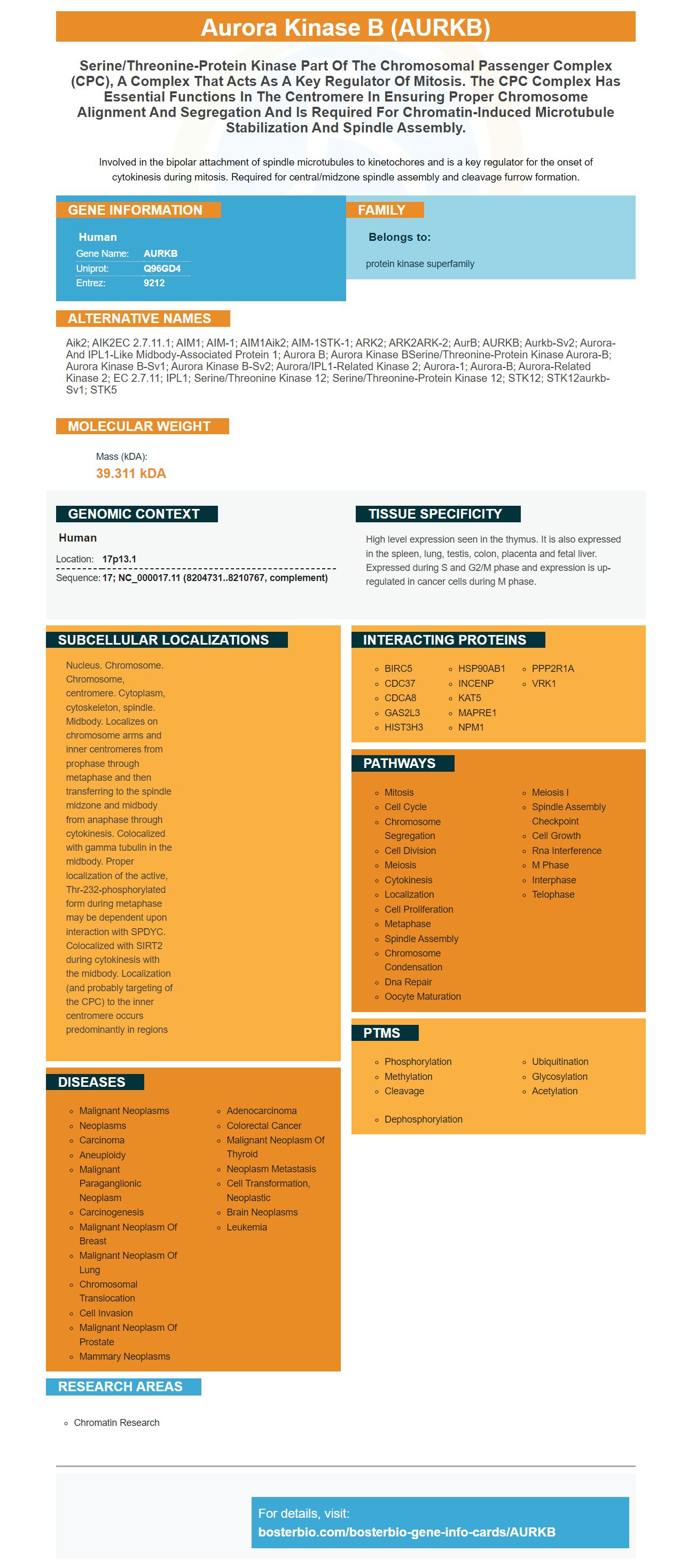
Facts about Aurora kinase B.
Involved in the bipolar attachment of spindle microtubules to kinetochores and is a key regulator for the onset of cytokinesis during mitosis. Required for central/midzone spindle assembly and cleavage furrow formation.
Human | |
---|---|
Gene Name: | AURKB |
Uniprot: | Q96GD4 |
Entrez: | 9212 |
Belongs to: |
---|
protein kinase superfamily |
Aik2; AIK2EC 2.7.11.1; AIM1; AIM-1; AIM1Aik2; AIM-1STK-1; ARK2; ARK2ARK-2; AurB; AURKB; aurkb-sv2; Aurora- and IPL1-like midbody-associated protein 1; Aurora B; aurora kinase BSerine/threonine-protein kinase aurora-B; aurora kinase B-Sv1; aurora kinase B-Sv2; Aurora/IPL1-related kinase 2; aurora-1; aurora-B; Aurora-related kinase 2; EC 2.7.11; IPL1; serine/threonine kinase 12; serine/threonine-protein kinase 12; STK12; STK12aurkb-sv1; STK5
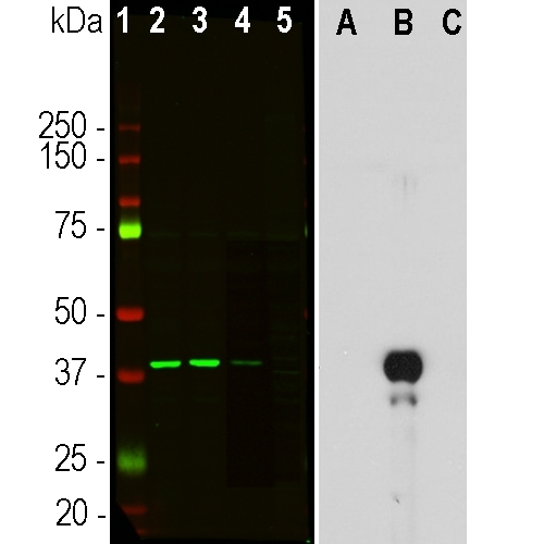
Mass (kDA):
39.311 kDA
Human | |
---|---|
Location: | 17p13.1 |
Sequence: | 17; NC_000017.11 (8204731..8210767, complement) |
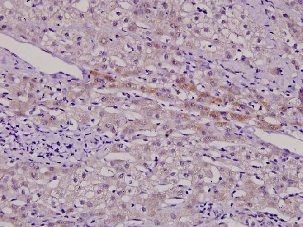
High level expression seen in the thymus. It is also expressed in the spleen, lung, testis, colon, placenta and fetal liver. Expressed during S and G2/M phase and expression is up-regulated in cancer cells during M phase.
Nucleus. Chromosome. Chromosome, centromere. Cytoplasm, cytoskeleton, spindle. Midbody. Localizes on chromosome arms and inner centromeres from prophase through metaphase and then transferring to the spindle midzone and midbody from anaphase through cytokinesis. Colocalized with gamma tubulin in the midbody. Proper localization of the active, Thr-232-phosphorylated form during metaphase may be dependent upon interaction with SPDYC. Colocalized with SIRT2 during cytokinesis with the midbody. Localization (and probably targeting of the CPC) to the inner centromere occurs predominantly in regions
If you are in the market for the best antibody for molecular biology, you should consider getting AURKB Marker. The company was established back in 1993. They have been producing signature antibody products and ELISA tests ever since. Recently, they have also expanded their product portfolio to include PCR-related molecular biology products. Boster Bio can likely offer something for all research needs. They offer technical support free of charge, 24 hour support by phone, and a wide range of other services to scientists.
Boster Bio's high affinity polyclonal antibodies are a great option if your search is for a brand new antibody. Boster's products are highly regarded by the research community, and have been cited multiple times over the last 25 years. They have also been validated for use in Western Blotting, Immunohistochemistry, and ELISA. They provide researchers high-quality antibodies that can be used in a variety of applications.
In immunosensor-based pathogen detection systems that use polyclonal antibodies, their higher avidity means they can stabilize the antigen-antibody complex longer. Polyclonal antibodies are more stable than monoclonal. They have multiple interactions that keep the antibody in the lattice. These interactions result is a higher immunofluorescence. Boster Bio high-affinity, polyclonal antibodies with superior performance are also a great choice for application testing.
Using a method of affinity chromatography, the purified pAbs are separated from their light chains by reducing their concentration. Kd is the proportion of the light and heavy chains. The eluted IgGs will be weighed and stored at 20 degrees Celsius until further analysis. Researchers can identify antigen-antibody interactions between cellular components using these antibodies for immunoglobulin M and IgG (IgG–HRP).
Polyclonal antibodies are more specific than monoclonal antibody. They recognize the same antigen and identify different epitopes on the target. In 1973, Dean and Abelseth used inactivated rabies virus (RABV)-infected mouse brain suspensions as antigens. The authors then obtained hyperimmune and specific antibodies in high concentrations.
These ELISA kits can be used to quantitatively determine low picogram levels (or plasma) of the human connecting peptide. This test can only be used for research. The half-life human C-peptide is about three to fourfolds that of insulin. It is therefore a useful marker for beta cell production. AURKB is just one of many phosphorylation markers.
Boster Bio has a wide range of high-quality antibodies for IHC-optimized research. We also answer some of the common questions that our customers ask us about our products. You can read on to learn more about how we make our IHC product as strong and durable as possible. You can also visit our FAQs to find the answers to your most common questions.
The AURKB marker is widely used in a variety of biological assays, including cell culture, tissue culture, and cancer research. The marker binds with the phosphorylation location of a downstream enzyme. The antibody can bind to casein and histone H3, as well as TP53. The assay results will show a single- or multiple-band signal. The AURKB marker has many applications, including detecting the presence of DNA and RNA.
Boster Bio's gene informationgraphics provide basic information about each gene. The infographics contain basic information on all genes, both human and mouse. A search bar makes it easy for you to find the gene that interests you. Boster's gene infographics can help you learn more about genes and the ways they regulate our bodies. The information can be used by you to learn more about your genes and to make your treatments more personalized. Read on to find out more about these handy tools.
PMID: 9514916 by Shindo M., et al. cDNA cloning, expression, subcellular localization, and chromosomal assignment of mammalian aurora homologues, aurora-related kinase (ARK) 1 and 2.
PMID: 9809983 by Tatsuka M., et al. Multinuclearity and increased ploidy caused by overexpression of the aurora- and Ipl1-like midbody-associated protein mitotic kinase in human cancer cells.