This website uses cookies to ensure you get the best experience on our website.
- Table of Contents

1 Citations

Facts about Copper-transporting ATPase 2.

.
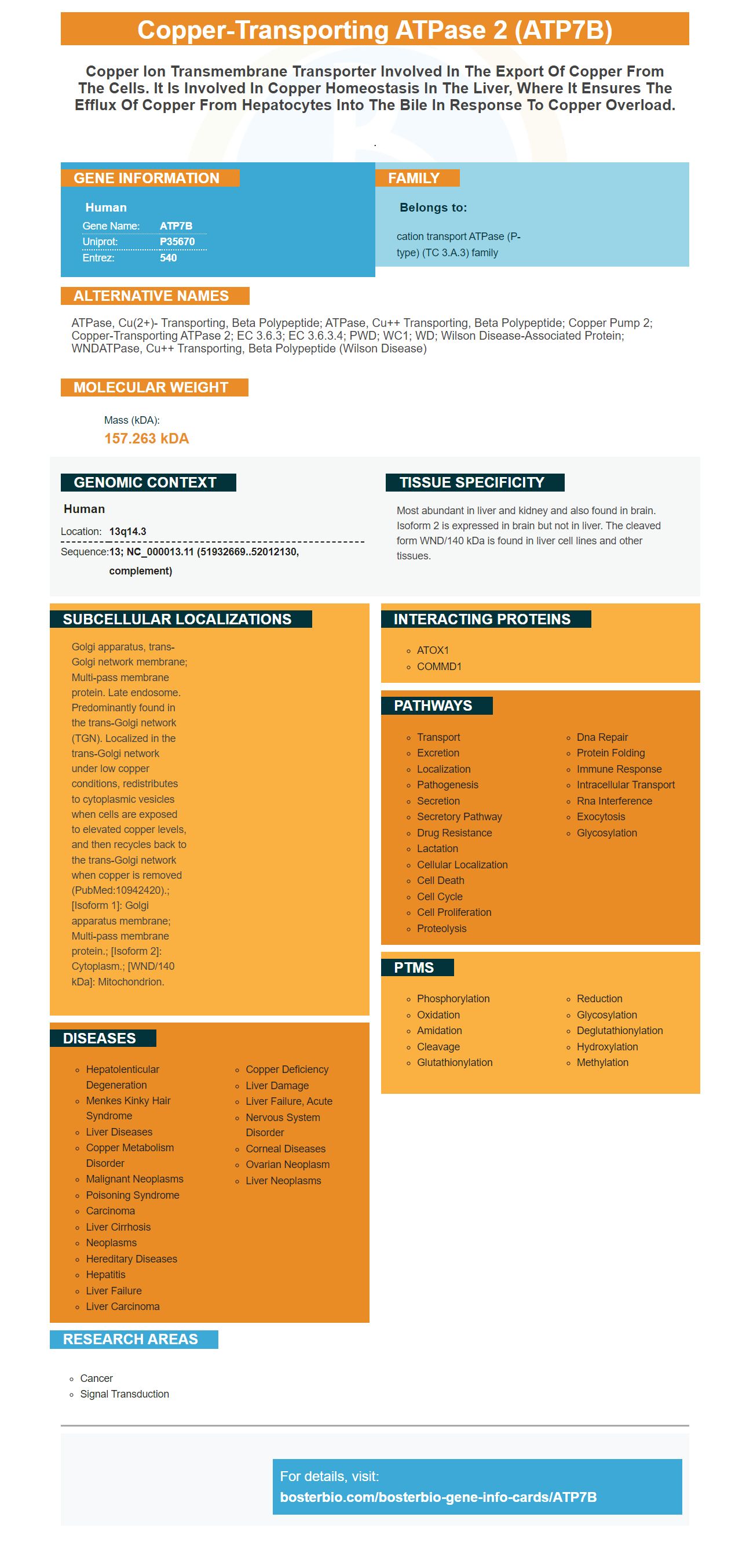
| Human | |
|---|---|
| Gene Name: | ATP7B |
| Uniprot: | P35670 |
| Entrez: | 540 |

| Belongs to: |
|---|
| cation transport ATPase (P-type) (TC 3.A.3) family |

ATPase, Cu(2+)- transporting, beta polypeptide; ATPase, Cu++ transporting, beta polypeptide; Copper pump 2; copper-transporting ATPase 2; EC 3.6.3; EC 3.6.3.4; PWD; WC1; WD; Wilson disease-associated protein; WNDATPase, Cu++ transporting, beta polypeptide (Wilson disease)
Mass (kDA):
157.263 kDA

| Human | |
|---|---|
| Location: | 13q14.3 |
| Sequence: | 13; NC_000013.11 (51932669..52012130, complement) |
Most abundant in liver and kidney and also found in brain. Isoform 2 is expressed in brain but not in liver. The cleaved form WND/140 kDa is found in liver cell lines and other tissues.
Golgi apparatus, trans-Golgi network membrane; Multi-pass membrane protein. Late endosome. Predominantly found in the trans-Golgi network (TGN). Localized in the trans-Golgi network under low copper conditions, redistributes to cytoplasmic vesicles when cells are exposed to elevated copper levels, and then recycles back to the trans-Golgi network when copper is removed (PubMed:10942420).; [Isoform 1]: Golgi apparatus membrane; Multi-pass membrane protein.; [Isoform 2]: Cytoplasm.; [WND/140 kDa]: Mitochondrion.


As a primary antibody provider, Boster offers high-affinity primary antibodies validated on Western Blotting, Immunohistochemistry, and ELISA. Over the past 25 years, Boster antibodies have been proven reliable and effective. We have published detailed protocol instructions to ensure the highest quality ATP7B testing results. Continue reading to learn the best uses of ATP7B peptide.
ATP7B is also known as the copper-transporting ATPase. It is a member in the ATPase family and transports metals with energy stored within ATP. It is found mainly in the liver. ATP7B plays a role in copper transport and can be useful in removing excess metal from the body. It is still not clear what the exact mechanism of this protein is.
The ATP7B peptide concentrations were comparable between normal controls and carriers in the study. The ATP7B concentration in DBS samples was also within the range of normal controls and WD patients. These results indicate that the BosterBioATP7B1056 peptide remains stable in blood for a considerable time. NBS also has some advantages, but immuno-SRM is more reliable than NBS.
In 92.1% cases, ATP7B quantitative analysis was able identify WD patients. This method also reduced the ambiguity associated with Cp and genetic analyses. The proposed diagnostic score can be used as an addition to the Leipzig and Cp scoring system. This study may assist in the diagnosis and treatment of patients with WD. Further research is needed to validate the results.
Fit-for-purpose guidelines were used to evaluate assay performance. A five-point response curve, which measures linear dynamic range, was created. Each level of concentration was replicated three times, with three samples analyzed at each time point to account for protein extraction from the DBS card. The assay demonstrated a linear response with r2 = 0.99. It covers a concentration range of 27 to 16,765 Pmol/L. Results were considered unreliable if the S/N ratio exceeded ten.
Wilson disease patients can have up to 37% alleles because of the h2069Q gene mutation. Studies of the ATP7B cDNA have shown that a single copy restores cell viability and rescues the mottled phenotype. It can also be used to increase the synthesis ATP7B, which is essential in cell recycling.
Wilson's disease can be diagnosed by genetic studies of ATP7B. Ineffective management of the condition can be possible if there is delay in diagnosing it. The researchers devised a novel method to measure ATP7B protein levels using dried blood spots taken from patients. The study involved the collection of 264 samples with IRB approval. It was discovered that the protein concentrations in the dried blood spots of patients containing ATP7B were consistent with genetic and biological data.
The ATP7B marker is a sensitive marker that identifies MMP-9 in humans. It is highly specific and sensitive. The ATP7B is a natural marker that detects the presence of this protein inside cells. The platform uses two antibodies to identify this marker. One antibody recognizes ATP7B while the other detects EEAl. Cell Signaling Technology has the anti-EEAl antibody.
The area under the curve (AUC), of each ATP7B peptide was 0.98 in a study comparing the two ATP7B assays. The LLOD (low light toxicity) was determined by dividing AUC and variance. This resulted in a 91.2% LLOD for ATP7B samples derived from healthy controls. The immunoSRM assay is also superior to the NBS.
The ATP7B peptide concentration was determined in two sets of patients: one group had a Cp level of ten to twenty mg/dL, and the other two groups had a Cp of less than 20 mg/dL. 71 of these patients had a low ATP7B level, while the two other groups had normal Cp levels. The immuno-SRM was also capable of identifying patients with common mutations.
In the current study, a total of 216 patients were included, with a prevalence of approximately 16%. The diagnosis was deemed highly probable by the fact that 172 of 226 patients (or a majority) showed ATP7B peptide levels above the cutoff. The algorithm can be validated by mutation analysis if it is performed on a large cohort. These data will be helpful in establishing diagnosis and treatment for WD patients.
ATP7B, a common gene, has over 400 variants. These variants cause a founder phenomenon and are associated to severe liver disease. There are several different types of ATP7B, but the most common are p.R778L, p.P992L, and p.T935M. These are described below. They are both able to detect the ATP7B signal in both types.
While ATP7B - 887 is an excellent diagnostic marker for WD, its low sensitivity may prevent a definitive diagnosis. In addition, it may not be sensitive sufficient to detect all affected individuals, particularly if the mutation is in the structure of the protein. To increase specificity and sensitivity, additional markers may need to be used. The ATP7B marker could be used as a screening marker for WD.
Wilson disease can be diagnosed by measuring the ATP7B peptide directly from dried blood spots. These variants can be found in a variety patients with WD. The most common is p.R778L in Jiangxi province and the Chinese mainland. However, in other regions, the p.P992L variant is also present. A study of this ATP7B peptide in a number of WD patients also revealed regional-specificity.
The study involved 158 patients with NSCLC, who had received platinum-based chemotherapy. It also compared ATP7B expression with DNA from matched normal samples. The MAFs of ATP7BSNPs varied in range from 91.2 to 100%. Furthermore, the genotype distributions were consistent with Hardy-Weinberg equilibrium. The use of ATP7B markers as a prognostic tool may be useful in clinical practice to help doctors determine the best treatment options for patients with NSCLC.
PMID: 7833924 by Petrukhin K., et al. Characterization of the Wilson disease gene encoding a P-type copper transporting ATPase: genomic organization, alternative splicing, and structure/function predictions.
PMID: 10334941 by Oh W.J., et al. Cloning and characterization of the promoter region of the Wilson disease gene.
*More publications can be found for each product on its corresponding product page

