This website uses cookies to ensure you get the best experience on our website.
- Table of Contents
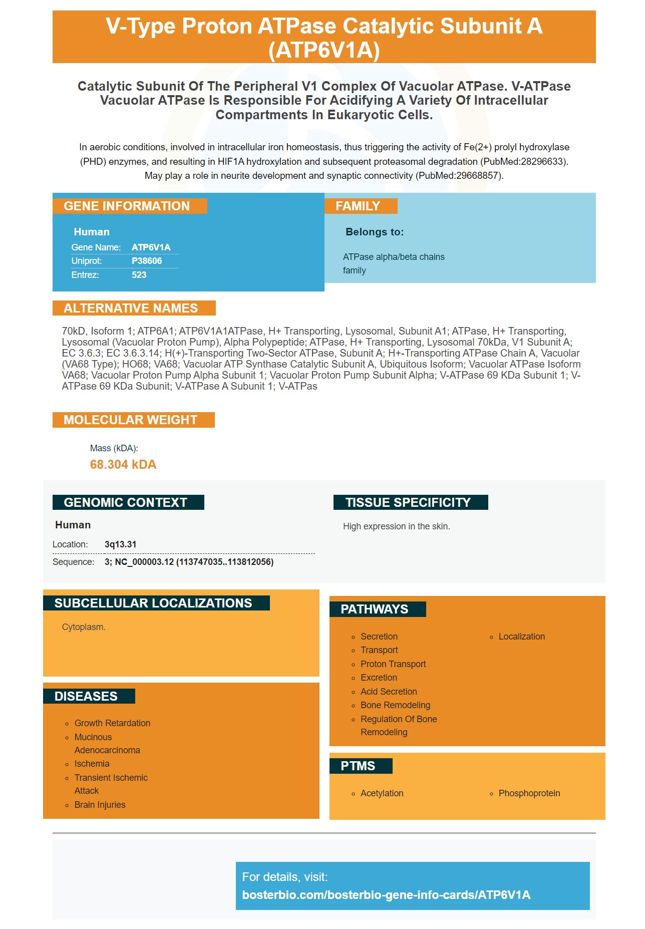


Facts about V-type proton ATPase catalytic subunit A.

In aerobic conditions, involved in intracellular iron homeostasis, thus triggering the activity of Fe(2+) prolyl hydroxylase (PHD) enzymes, and resulting in HIF1A hydroxylation and subsequent proteasomal degradation (PubMed:28296633). May play a role in neurite development and synaptic connectivity (PubMed:29668857).
| Human | |
|---|---|
| Gene Name: | ATP6V1A |
| Uniprot: | P38606 |
| Entrez: | 523 |

| Belongs to: |
|---|
| ATPase alpha/beta chains family |

70kD, isoform 1; ATP6A1; ATP6V1A1ATPase, H+ transporting, lysosomal, subunit A1; ATPase, H+ transporting, lysosomal (vacuolar proton pump), alpha polypeptide; ATPase, H+ transporting, lysosomal 70kDa, V1 subunit A; EC 3.6.3; EC 3.6.3.14; H(+)-transporting two-sector ATPase, subunit A; H+-transporting ATPase chain A, vacuolar (VA68 type); HO68; VA68; vacuolar ATP synthase catalytic subunit A, ubiquitous isoform; Vacuolar ATPase isoform VA68; vacuolar proton pump alpha subunit 1; Vacuolar proton pump subunit alpha; V-ATPase 69 kDa subunit 1; V-ATPase 69 kDa subunit; V-ATPase A subunit 1; V-ATPas
Mass (kDA):
68.304 kDA

| Human | |
|---|---|
| Location: | 3q13.31 |
| Sequence: | 3; NC_000003.12 (113747035..113812056) |
High expression in the skin.
Cytoplasm.


This article will help you to comprehend ATP6V1A protein better. This article provides ATP6V1A homology, ATP6V1A mutations, immunolabelling and co-localization with LC3B. It also explains more about ATP6V1A and its applications in the fields of biology and medicine. Boster's products are tested on multiple platforms , and with known positive and negative samples. Boster provides product credits to first-time reviewers. This program is open to all scientists worldwide.
The ATP6V1A gene encodes the A subunit of the v-ATPase, a protein essential for pH homeostasis in all eukaryotic cells. The vATPase is particularly important in the functioning of synapse. Recent whole exome studies have discovered de novo heterozygous variants in the ATP6V1A gene. This gene mutation could lead to v-ATPase related encephalopathy.
The DESIRE project has identified a fourth patient. The study has provided preliminary results for the three patients who were initially identified. The project team analyzed the genetic variants inherited with more than 1 percent in the 1000g ExAC and dbSNP databases. Sanger sequencing confirmed the mutations on the genomic DNA of both the parents and the probands. In addition, the patients' parents were confirmed by using twelve microsatellite markers.
The ATP6V1A homology model was developed using the structure of the S. cerevisiae v-ATPase in the state of rotation 1. The model also showed overlapping mutation sites with E. the hirae V-ATPase. This is why the homology model ATP6V1A is of high confidence. It is important to remember that E. Hirae's mutations sites are placed on the same model to pinpoint the exact location.
The activation of vATPase results in the acidification of lysosomal cells. Inhibition of this process can inhibit active cell growth. Mutated ATP6V1B2-mutant ATP causes slight impairment in lysosomal acidsification. Mutated ATP6V1B2-mutant protein causes decreased degradation of lysosomal protein and an increase in autophagic flux.
Contrary to FL homozygous mutants, the variant known as ATP6V1B2 within the ATP6V1B2 genes mimics heterozygous FL cell mutation. The R381Q mutation in mammalian cells increases autophagy activity. It is identical to the ATP6V1B2 mutation in mammalian cells. In turn, the mutant FL cell is more sensitive to agents that induce autophagy.
The ATP6V1A homology model is based on two distinct mutants that confer a defect in the v-ATPase enzyme that is central to proton pump activity. The cells of HEK293T were injected with a pH-sensitive dual reporter dye that was subsequently bonded to dextran. The cells were then incubated using buffers of specified pH levels. This allowed a quantitative analysis of the mutants.
The ATP6V1A-related brain encephalopathy is an exciting new concept in the field of lysosomal diseases, with an essential role for the endo-lysosomal membrane protein, which is in a state of. These mutations result in altered pH homeostasisand intracellular accumulation of substrates, and alterations in the ATP6V1A proteins. These mutations cause neurodegenerative and developmental abnormalities in brain development and can often be identified during the prenatal period. The severity of the disease is dependent on the pathogenic variants that are responsible for the mutation.
D100Y is the most common variant that causes PDS. This variant alters the function of the lysosomal system and is associated with impaired synaptic development and synaptic connectivity. These issues could be involved in the pathophysiology of the pigmentation, PDS, and glaucoma. The future studies on this mutation and other diseases could provide more information on the underlying causes of phenotypes.
The D204 - R204 mutation in Nef reduced the effectiveness of mutant HIV-1. The mutations reduced the infectivity of mutant SIVmac239 in a recent study. Mutations in the ATP6V1Adomain caused a decreased infectivity for a mutant HIV-1. These results are consistent with those observed in human clinical studies.
The ATP6V1A protein encodes for the proton enzyme V-type ATPase catalyst subunit A. It is an enzyme with multiple subunits that regulates the acidification of organelles within the intracellular body. Organelle acidification is essential for the sorting of proteins. The enzyme is cytosolic and comprises a V1 domain as well as the transmembrane V0 domain. The V1 domain consists of the A and B subunits, as well as the C, D, E F, H, and D subunits. Each of these subunits contains an enzyme catalytic site for ATP.
The ATP6V1B2 gene encodes the V1B2 subunit of the V-ATPase enzyme, a proton pump responsible for acidification of lysosomes. Although it is not able to identify the underlying mechanisms the mutations in this gene can lead to problems such as DOORS and DDOD. Mutant mice with this gene are affected in a variety of areas such as the auditory system. While these findings suggest that ATP6V1B2 is responsible for auditory dysfunction in mice, the exact mechanism that the disorder manifests itself is still unknown.
The ATP6V1B2 subunit is connected to the ability of lysosomes to be acidified and is affected by the pH of the lysosome. A low pH could lead to the degradation of macromolecules and lysosomal storage issues. Additionally, the absence in this V-ATPase Subunit in human cells can inhibit the autophagy pathway and lead to the accumulation of nonfunctional Lysosomes.
ATP6ap2 is involved in receptor-mediated endocytosis, as well as lysosomal activity in the proximal tubule. Mutations in the ATP6ap2 gene hinder endocytosis and vesicular trafficking in mice. In addition, mice with knockouts do not have ATP6ap2 and show impairment in proximal tubule function.
This gene is involved with the recognition of many proteins. Cubilin is a partner in the ATP6ap2 gene. In the absence of this gene cubilin doesn't recognize any protein. Mice with mutations have lower levels of both proteins. Moreover, the gene expression of the ATP6ap2 receptor has been downregulated in Flox/Pax8 mice.
A positive new born of a ShAtp6ap2 transgenic line was used to establish the transgenic lines on SD background. The negative littermates served as wild type controls. These results support the theory that ATP6V1B2 co-localizes with LC3B in Boster Bio. This gene is therefore involved in the biochemistry and co-localization of the ATP6V1B2-LC3B.
PMID: 8463241 by van Hille B., et al. Identification of two subunit A isoforms of the vacuolar H(+)-ATPase in human osteoclastoma.
PMID: 28065471 by Van Damme T., et al. Mutations in ATP6V1E1 or ATP6V1A cause autosomal-recessive cutis laxa.


