This website uses cookies to ensure you get the best experience on our website.
- Table of Contents
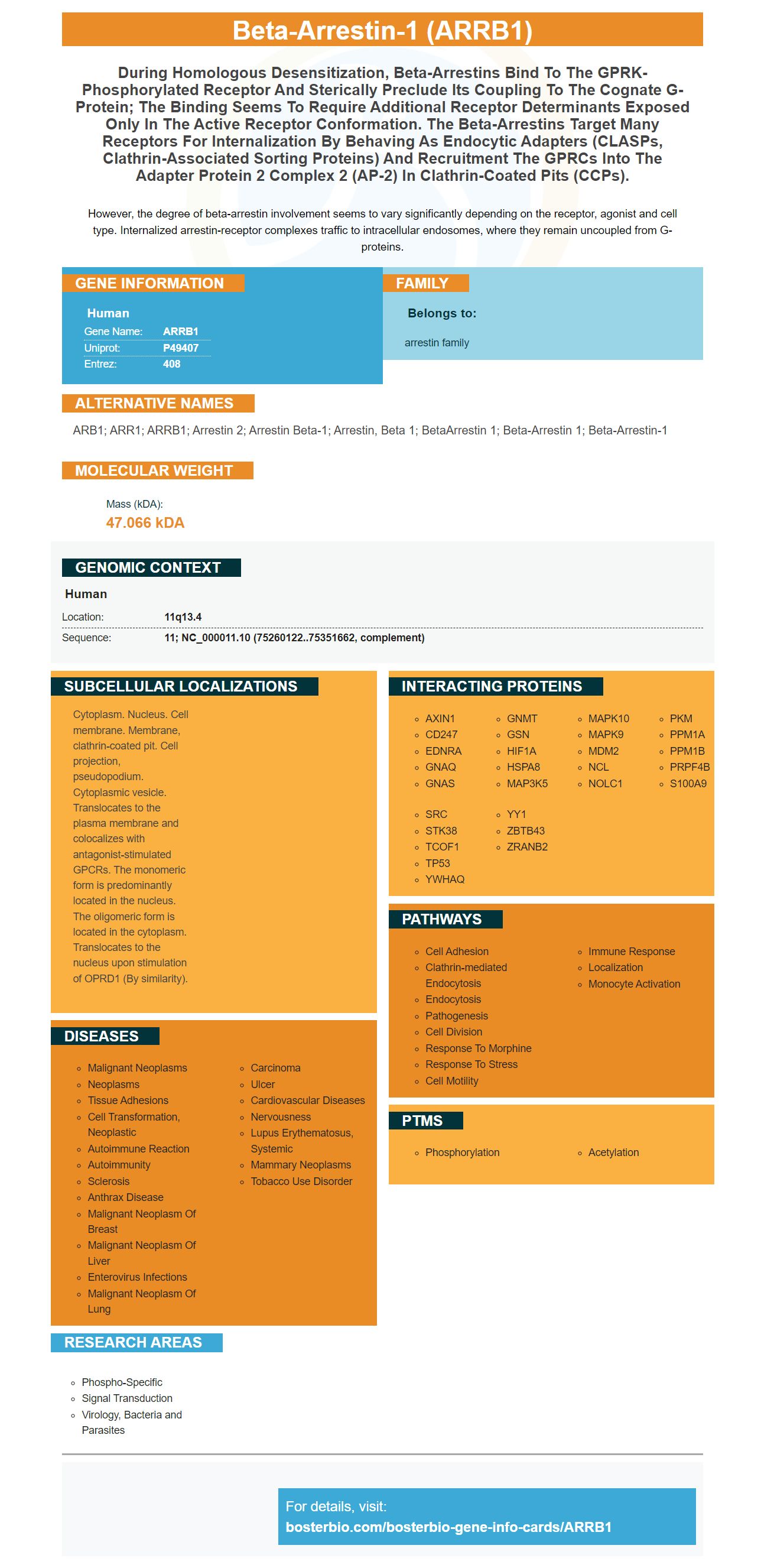


Facts about Beta-arrestin-1.

However, the degree of beta-arrestin involvement seems to vary significantly depending on the receptor, agonist and cell type. Internalized arrestin-receptor complexes traffic to intracellular endosomes, where they remain uncoupled from G-proteins.
| Human | |
|---|---|
| Gene Name: | ARRB1 |
| Uniprot: | P49407 |
| Entrez: | 408 |

| Belongs to: |
|---|
| arrestin family |

ARB1; ARR1; ARRB1; arrestin 2; Arrestin beta-1; arrestin, beta 1; betaArrestin 1; beta-Arrestin 1; beta-arrestin-1
Mass (kDA):
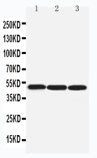
47.066 kDA

| Human | |
|---|---|
| Location: | 11q13.4 |
| Sequence: | 11; NC_000011.10 (75260122..75351662, complement) |
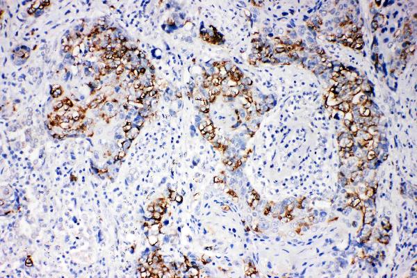
Cytoplasm. Nucleus. Cell membrane. Membrane, clathrin-coated pit. Cell projection, pseudopodium. Cytoplasmic vesicle. Translocates to the plasma membrane and colocalizes with antagonist-stimulated GPCRs. The monomeric form is predominantly located in the nucleus. The oligomeric form is located in the cytoplasm. Translocates to the nucleus upon stimulation of OPRD1 (By similarity).


If you're interested in finding the best ARRB1 ELISA kits, you've come to the right place. This page contains information about ARRB1's ubiquitination as well overexpression and plasmids. You can also learn more about the ELISA Ub plasmid, and the benefits of Picokine(tm).
Both the ARRB1 (or ARRB2) markers are essential to normal brain function. They also play a variety of roles in central nervous systems diseases. Both ARRB1 (and ARRB2) markers are involved inflammatory reactions. Both genes regulate trafficking and signaling. These receptors can also function as cellular transcript factors that mediate a healthy immune response. ARRBs also regulate mTOR complex 1 signaling.
ARRB1 & ARRB2 markers display distinct expression patterns depending on the cellular and vivacity of the environment. In the EAE model mouse model, knockouts ARRB1/ARRB2 decrease the severity, while the corresponding effects of ARRB1/ARRB2 on the inflammatory responses are more evident. ERK1/2 activation can be increased by downregulation of one marker, and decreased activation of the other. ARRB1 and ARRB2 receptors also have opposite effects on NLRP3 inflammasome.
ARRB1 & ARRB2 play different roles in the pathogenesis and progression of PD. Both markers play an important role in microglia-mediated inflammation. Their opposite actions could help to develop drugs for PD. ARRB1 is an ARRB1 marker that can be used for research in this field. It can be used for identifying disease-related pathways and identifying potential treatment targets. Boster Bio is excited to offer this marker.
We used the Boster bioassay TGCT-FLS tissues to investigate the effects of Arrb2 omission in vivo. Both tissues showed high levels of Arrb2 and were not significantly different from the OA samples. Moreover Arrb2 had a positive correlation with FLS proliferation. Therefore, we chose these cells as our model for further studies. These findings could be misleading. We recommend consulting a qualified biologist or biotechnologist to complete the experiment.
The findings revealed that ARRBs play divergent roles in the pathogenesis of several central nervous system diseases. In addition, ARRB2 has recently been implicated in PD. It mediates the functions and k-opioid/dynorphin receptors, protects the TH positive neurons against inflammation, and plays an important role in the development L-DOPA dyskinesia.
Boster Bio's ARRB2-Ubplasmid contains two DNA fragments encoding ARRB2 and ubiquitin. The ARRB2-Ub protein itself is encoded by a 1227-bp DNA length. These plasmids are encoding mutant ARRB proteins. They were created using site-directed mutation kits. DNA sequencing confirmed that mutations were present. The mutations are compatible and compatible with the wild type protein.
The knock-outs of ARRB1 and ARRB2 had opposite effects on DA neuron loss, microglia activation, and neuroinflammation. The results of these experiments provide novel insights into the functional divergence of ARRBs in PD. These plasmids are easy to use and have a simple vector system. In a separate experiment, we used a plasmid containing a promoter specific to microglia to knock down ARRB1 & ARRB2 respectively in a rat model PD.
ARRB1 is a novel gene with diverse functions in various tissues and cells. It interacts with three NFKB pathway molecules, including p65. These interactions vary based on the level of inflammatory stimulation. Knockout of any ARRB isoforms increased the interaction with p65. This interaction has important implications in understanding the mechanisms that underlie human cancer and other diseases.
This study found that the ARRB-mediated regulation of microglia inflammation was regulated by the p65 protein. We then used RNA-seq to normalize the gene expression levels of pro-inflammatory markers. We detected differential expression in 15 genes using Arrb2-/ mice that had been treated with LPS plus IFN g for 6 hours. The log2-fold changes in expression were the result.
ARRB1 can affect a variety of cellular systems and animals, according to molecular analyses. It has been shown that Arrb1 knockouts improve EAE symptoms, while ARRB2-/ mice have more severe EAE disease. ARRB1-/ mice have higher levels of Il1b and Tnf.
Despite its close relationship to STAT1 & NF kB, Nprl3 plays opposing roles in microglia-mediated inflammation & PD. This finding offers new insights into the functional divergence between ARRBs and PD. The results show that Nprl3 also controls the expression and activation STAT1/p65.
Dopaminergic neuronal protection in MPTP/p mice is achieved by knockdown Nprl3. The knockdown protects dopaminergic neuron in this mouse model for Parkinson's disease. It also reduces the inflammation in the model. These findings suggest that the Nprl3 inflammasome may play an important role in the development of Parkinson's disease.
ARRB1 regulates Nprl3, which can be expressed in microglia. ARRBs activate microglia-specific inflammation signals. Studies using primary cell cultures have shown that ARRB1 and ARRB2 are important in controlling inflammation in microglia. These findings provide important insight into the role that ARRBs play in PD.
Boster Bio has created several ELISA kits to detect ARRB1 under a variety of pathological or physiological conditions. It includes more than 12,000 antibody samples that have been validated to detect ARRB1 in WB, IHC, and Flow. Boster Bio antibodies have been tested for specificity and affinity against 250 tissue panels. They are also quantitatively validated against known amounts of recombinant proteins. Boster Bio's immunological substances are available at tebu.bio
Many studies have shown that ARRBs regulate several pro-inflammatory genes. ARRB1/ mice have higher expression of the Nos2, Tnf and Il1b markers. This suggests that these two proteins may have opposite effects upon the inflammatory response. These genes regulate each other and play a variety of biological functions. Arrb2 plays a significant role in neuronal growth and is an important component the mTOR complex.
The ARRB1 marker is versatile and can be used in many immunological applications. Boster Bio provides ELISA kit and research antibodies for the detection biomarkers. These include cancer, neurosciences (inflammation), and other areas. These antibodies have a high sensitivity, with sensitivity of up to one picogram. In addition, they are compatible with a variety of detection methods, including IHC and Flow Cytometry. Boster Bio products are available from tebu-bio, including in-house validation of ELISA kits.
ARRB1 has been shown to regulate the NFKB signaling pathway. It also regulates the STAT1 signaling pathways and MAPK signaling pathways. The ARRB1 and ARRB2 markers can be used to study the interactions between the NF-kB pathway. ARRB1 and ARRB2 are known to interact with p65 in various co-IP assays. Knockouts one ARRB Isoform increased interaction with p65.
ARRB1 targets include those that relate to cell cycle, such as nucleosome organisation or protein/DNA complex assembly. ARRB1 targets are mostly associated with cell metabolism or cell cycle. ARRB1's specific applications have only been reported to date. ARRB1 is a useful tool to study gene regulation in cell biology.
The Warburg effect, first described by Otto Warburg in the 1920s, is a common feature of cancer cells. Normal cells rely mainly on oxidative phosphorylation for growth, but tumour cells rely more heavily on aerobic glycolysis to fuel cell proliferation. This metabolic shift is caused by the ARRB1 Nuclear Protein. It regulates HIF1A transcriptional activities and the expressions glycolytic gene products.
One study revealed that ARRB1 interacts directly and can enhance transcription activity by interacting with the hypoxia inducible factor 1A protein (HIF1A). HIF1A is then hydroxylated at specific proline residues by PHDs, tagged for ubiquitination, and degraded by the proteasome pathway. HIF1A, ARRB1, and ARRB1 have the same function but target different targets and promote various types of tumour growth.
PMID: 8486659 by Parruti G., et al. Molecular analysis of human beta-arrestin-1: cloning, tissue distribution, and regulation of expression. Identification of two isoforms generated by alternative splicing.
PMID: 9501202 by Aragay A.M., et al. Monocyte chemoattractant protein-1-induced CCR2B receptor desensitization mediated by the G protein-coupled receptor kinase 2.