This website uses cookies to ensure you get the best experience on our website.
- Table of Contents
9 Citations 18 Q&As
5 Citations 16 Q&As
3 Citations
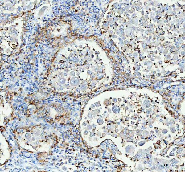
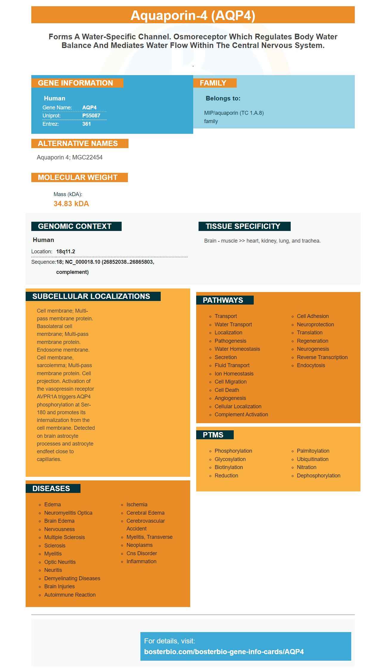
Facts about Aquaporin-4.
.
Human | |
---|---|
Gene Name: | AQP4 |
Uniprot: | P55087 |
Entrez: | 361 |
Belongs to: |
---|
MIP/aquaporin (TC 1.A.8) family |
aquaporin 4; MGC22454
Mass (kDA):
34.83 kDA
Human | |
---|---|
Location: | 18q11.2 |
Sequence: | 18; NC_000018.10 (26852038..26865803, complement) |
Brain - muscle >> heart, kidney, lung, and trachea.
Cell membrane; Multi-pass membrane protein. Basolateral cell membrane; Multi-pass membrane protein. Endosome membrane. Cell membrane, sarcolemma; Multi-pass membrane protein. Cell projection. Activation of the vasopressin receptor AVPR1A triggers AQP4 phosphorylation at Ser-180 and promotes its internalization from the cell membrane. Detected on brain astrocyte processes and astrocyte endfeet close to capillaries.
When you're ready to explore your protein of interest, Boster Bio's primary-secondary-ABC system and Super Vision Detection kits are a great choice. Boster Bio's AQP4 marker can help you identify proteins with low background or high specificity. Continue reading to find out more about the AQP4 marker and its many benefits.
The primary-secondary-ABC system uses avidins conjugated to signal molecules, usually Horseradish Peroxide (HRP), to identify proteins of interest. It is highly sensitive and has low background. Other detection systems have been developed with organic polymers and polysaccharides, such as Boster Bio's SuperVision Detection Kits.
The products offered by the company include a range of high-quality cell lines as well as reagents. In addition, it offers cell, biosample and histology services. The company also produces TALEN AAV, lentiviral, and TALEN systems. It also provides Proteomics and genomics products. Its collection of reagents include TALEN-mutagenesis, lentiviral RNA, and different antibodies.
With the Boster Bio SuperVision detection kits, researchers can easily locate proteins in complex samples with minimal background. The ELISA kits can be used with a variety of samples. Validation images and procedures for the kits can be obtained on request. These kits utilize Picokine(tm) technology to increase the sensitivity of the test and reduce background, saving researchers more than 30 minutes in IHC. They also use the Picoband technology which incorporates information into the most effective immunogen design and optimization. Technical support is available and is backed by a vast knowledge base.
The AQP4 marker could be a tool in Alzheimer's research. This protein is a catalyst for elimination of waste in the glymphatic system. In healthy brains, AQP4 is predominantly expressed on astrocyte endfeet. CSF circulates through the para-arterial pathway into the brain parenchyma and veins. Sadly, with aging, AQP4 depolarization and polarization decline. In AD the glymphatic system is less efficient and a build-up of beta-amyloid takes place.
AQP4-IgG antibodies can detect the development of NMOSD , and allow the faster treatment. AQP4IgG antibodies can be identified with high sensitivity and specificity by using an in-vivo test. Human embryonic kidney cells were transfected with M23 AqP4 that was fluorochrome-tagged for this assay. The cells were then incubated with human serum. The co-localization of human IgG and AQP4 was then evaluated.
There are a variety of tissues that express AQP4 in their neurons. It is usually found on the astrocyte that is closest to a blood vessel. In some instances however, AQP4 is mislocalized and does not target the blood vessels at the end of the foot. Studies on rats have indicated that AQP4 regulates glutamate levels and the expression of GLT-1.
There are numerous other advantages to the AQP4 marker. It is used extensively in the astrocyte-derived cell culture. It enhances water permeability and cell adhesion. Anti-AQP4 antibodies target this particular protein. They could also detect neuron-glial interactions in retinas which are distorted due to an AQP4 knockout.
It is also associated with an autoimmune disorder, called NMOSD. This condition is characterized by the loss of astrocytes, and the demyelination of the optic nerve and spinal cord. Most patients with NMOSD have demyelinating lesions spanning three or more vertebral segments and more than 80% patients are found to have autoantibodies to the protein. A rodent study revealed a pattern of perivascular binding for the AQP4 antibodies.
While numerous studies have revealed that AQP4's IgG-mediated CDC requires human complement for pathogenicity there are many potential benefits of this antibody. Anti-AQP4IgG-IgG binds with astrocyte stimulate glial cells that express FcR. The anti-AQP4IgG-IgG treatment produced less pathology than wild-type AQP4IgG.
The AQP4–IgG–IgG–IgG serology increased sensitivity and specificity in CSF but also has other benefits. AQP4-IgG-IgG immunoadsorption may reduce the complement system and decrease the titer of blood serum. This, in turn, may reduce the risk of developing cancer and other illnesses.
AQP4 is found in neural tissues. It may aid in the elimination of beta peptides from amyloid and regulate extracellular space volume during synaptic activity. In the retina, AQP4 is expressed in Muller glial cells. The Muller glial cells support structure in the retina, maintain the microenvironment of the retina, and regulate neuronal activity. The vision function of the retina is assessed by electroretinograms.
PMID: 7559426 by Yang B., et al. cDNA cloning, gene organization, and chromosomal localization of a human mercurial insensitive water channel. Evidence for distinct transcriptional units.
PMID: 8601457 by Misaka T., et al. A water channel closely related to rat brain aquaporin 4 is expressed in acid- and pepsinogen-secretory cells of human stomach.
*More publications can be found for each product on its corresponding product page