This website uses cookies to ensure you get the best experience on our website.
- Table of Contents
2 Citations 6 Q&As
1 Citations 5 Q&As
Facts about Aquaporin-3.
Provides kidney medullary collecting duct with high permeability to water, thereby permitting water to move in the direction of an osmotic gradient. Slightly permeable to urea and may be a water and urea exit mechanism in antidiuresis in collecting duct cells.
Human | |
---|---|
Gene Name: | AQP3 |
Uniprot: | Q92482 |
Entrez: | 360 |
Belongs to: |
---|
MIP/aquaporin (TC 1.A.8) family |
AQP-3; aquaglyceroporin-3; aquaporin 3 (GIL blood group); aquaporin 3 (Gill blood group); aquaporin 3; aquaporin-3; GIL; Gill blood group
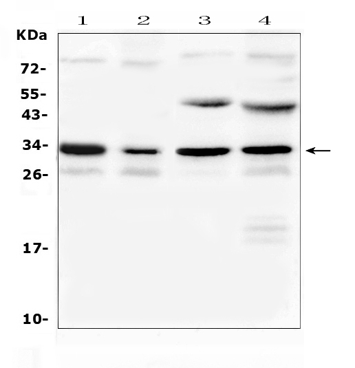
Mass (kDA):
31.544 kDA
Human | |
---|---|
Location: | 9p13.3 |
Sequence: | 9; NC_000009.12 (33441154..33447596, complement) |
Widely expressed in epithelial cells of kidney (collecting ducts) and airways, in keratinocytes, immature dendritic cells and erythrocytes. Isoform 2 is not detectable in erythrocytes at the protein level.
Cell membrane; Multi-pass membrane protein. Basolateral cell membrane; Multi-pass membrane protein.
The AQP3 marker is expressed by various malignant tumor cells. It regulates the expression of CD133 and also the regulation of invasion and migration of HTR8/Svneo cell. It can also affect human reproduction. This is a summary of its most effective uses. Hope you can gain from this new information. Boster Bio: The Most Effective Uses of the AQP3 Marker
Recent studies have shown that hypoxia can increase AQP3, which is upregulated in hypoxic HCC cells. Hypoxia-prone HCC cells are less sensitive than normal to sorafenib. This drug blocks the growth and death of cancer cells. It is unclear what significance AQP3 is playing in the hypoxic growth of HCC cells.
The AQP expression is related to tumor grade in different cancer types which includes astrocytoma. In addition, tumor cells with higher AQP expression were more likely to be in an advanced disease stage. Furthermore, expression of AQP 9 was detected in malignant ovarian tissues and this correlated with tumor grade. The inhibition of AQP-1 slowed the spread of lung cancer cells which decreased tumor grade.
AQP 3 is a protein that functions in the extracellular matrix by acting as a mediator for cytoskeleton rearrangement of actin and helping to facilitate cell migration. It also regulates the expression of mesenchymal markers. Cao and. al. discovered that FGF-2 induces AQP3 expression and that cells that express shRNA with inhibitory AQP3 reduced the movement of cells induced by FGF-2 by 50 percent. Moreover, AQP3 is associated with activation of the ERK1/2 and Akt pathways.
Bioinformatics analysis was used to determine the AQP3 mRNA levels and protein levels in various cell varieties of HCC. Using RNAIsoPlus assay kit, total RNA was extracted and converted to cDNA. After that, SYBR-Green master mix for qPCR was used to determine the presence of the targeted gene. The results showed that AQP3 was overexpressed in HCC cells by 33.5 percent in comparison to control cells.
Moreover, overexpression of AQPs has been associated with better survival rates in cancer of the stomach, especially in women , and in early-stage tumors. Further research into AQPs activity could offer an universal marker for gastric malignancy. Although the prognostic significance of specific AQPs proteins is not yet determined, it may be a factor when targeted treatments are developed for gastric cancer.
AQP3 is a factor that affects the keratinocyte's cell migration. AQP3 decreases activity of p38 MAPK which is a cell-migration signaling protein. The overexpression of AQP3 can lead to the formation of L-cell cell cluster formation. The gene is expressed in a variety of malignant tumor cells and its expression in these cells suggests that it is linked to the development of cancer.
Recent studies have shown that the AQP3 marker can promote the transcription of CD133, a gene that has a role in the development of HCC. It has been shown that CD133 is a key factor in HCC growth, invasion, self-renewal, and self-renewal. Its expression may also be used to predict the growth of HCC by acting as a marker. However, the molecular basis for AQP3-regulated CD133 expression remains unclear.
Our research revealed that AQP3 expression was correlated with CD133. This was further supported by the association of AQP3 and CD133. In addition CD133 and AQP3 were found to be positively correlated in a panel of 86 markers that are involved in cell proliferative and metastasis. These results suggested that AQP3 may play a role in the process of tumorigenesis.
AQP3 activates the signaling pathways of b-catenin by activating Wnt GSK-3b and human GC cells. We first determined whether AQP3 could activate the signaling pathways for b-catenin in CD133+ cells by controlling Wnt's expression and reducing its GSK-3b activity. To reduce CD133 levels of mRNA as well as protein, we transfected cells with Lv–AQP3-shRNAs. Additionally, we used double immunofluorescence in order to determine whether CD133 and AQP3 colocalized in these cells.
AQP3 also stimulates transcription of CD44 which is essential in stem cell formation. This protein activates the Wnt/GSK-3b pathway which allows stem-like characteristics to be obtained in GC cells. These data suggest that the AQP3-mediated transposition of CD133 in hepatitis C cell lines might be in a position to predict resistance to treatment.
To confirm that AQP3 regulates CD133 expression in HCCLM3 cells, we first used lentivirus-AQP3-shRNA to transduce HCCLM3 cells or Huh7 cells. Cells were treated with lentivirus-AQP3-shRNA or Lv-AQP3-NC for 48 hours. The expression of AQP3 was then analyzed by QRTPCR. We also confirmed that AQP3 expression is regulated by a cell cycle.
The AQP3 marker enhances the expression of CD44 in the GC cells. Researchers have previously shown that AQP3 can boost the expression of CD44 within the GC cells. This is associated with an increased chance of disease progression. AQP3 can also increase the activity in the nucleus of bcatenin. Additionally, AQP3 may regulate the expression of CD44 in GC cells.
AQP3 is extensively expressed in human IBC cells. AQP3 and AQP5 are closely connected in lung cancer. It is believed that it helps regulate the growth and expansion of these cells. AQP3 has been proven to regulate tumor osmoregulation. It is also believed to influence the growth of the HTR8/Svneo cell lines. It also regulates the inflammation response.
Hypoxia regulates important genes, including those that are involved in the development of astrocytes. Boster Bio's AQP3 marker is a key factor in the migration and invasion of Svneo/HTR8 cells. The results of this study are consistent with previous findings. Hypoxia can trigger transcription HIF which is involved in neuronal dysfunction as well as Ewing's Sarcoma. Hypoxia-induced HIF siRNAs are available for further research.
AQP3 regulates transport of hydrogen peroxide is involved in downstream signaling, and regulates the expression of EMT proteins. These properties of AQP3 suggest that it could be a therapeutic target. Furthermore, it is important to note that AQP3 is a key player in the transportation of glycerol in cells and promotes intracellular ATP.
The authors of the study included Koritzinsky, Marianne, Heun-Johnson, and Turner. They conducted experiments on cells in a controlled environment. The cells were able to move in physically restricted spaces despite the absence of myosin contraction or actin polymerization. This is in line with the hypothesis that AQP5 modulates cell volume.
Multiple tissues in the body contain AQPs. These changes could affect the development of various types of cancer and tumors. These substances can also be observed in human erythocytes. Thus, AQP3 expression should be of clinical significance in cancer research. So what are you wasting time for? Get the latest news from BosterBio and get on the speed-to-market.
A new study from Boster Bio has identified a new marker for HIV-1, which regulates the expression of two transcription factors for the T-box. This protein has been implicated in regulating the expression of HIV-1 Tat, which affects the response to psychostimulants and delay-dependent memories in mice. If this is indeed the situation, then the treatment for HIV-1 may be just as straightforward as fixing the root cause.
Aquaporin-3, also referred to as AQP3 is a crucial element in the regulation of glycerol levels in skin cells. The levels of AQP3's expression can be used to determine the presence of the gene. This is crucial for normal human reproduction. Boster Bio created antibodies against Aquaporin-3 with mouse and rabbit models. The AQP3 gene is expressed at higher levels in spermatozoa and its expression is significantly increased during the initial stages of the human reproductive process.
In mouse models, AQP3 was detected in the cell membrane of trophoblasts where it was associated with embryo development and adhesion to the endometrium. It is not known if AQP3 expression is detected in trophoblasts that are not derived from human pregnancy, or whether it plays a role in embryo implantation. The most effective uses of the AQP3 in human reproduction are in the process of being developed.
Researchers used gene expression microarrays to determine the pathways regulated during embryo implantation by AQP3. Any fold-change greater than 2 was considered to be significantly differentially expressed genes. For AQP3 downregulation 311 genes were shown to be significantly different. These genes were associated with angiogenesis, cell movement, extracellular matrix composition recombination and the inflammatory response.
Aquaporins, also called channel proteins, regulate the levels of water and small molecular compounds such as urea or nitrogen. Multiple AQPs have been discovered in embryos of humans prior to implantation. AQP3 is expressed from the zygote phase to the blastula stage. AQP3 is an important aquaporin in embryo development in human embryos.
PMID: 7558005 by Ishibashi K., et al. Structure and chromosomal localization of a human water channel (AQP3) gene.
PMID: 12239222 by Roudier N., et al. AQP3 deficiency in humans and the molecular basis of a novel blood group system, GIL.
*More publications can be found for each product on its corresponding product page