This website uses cookies to ensure you get the best experience on our website.
- Table of Contents
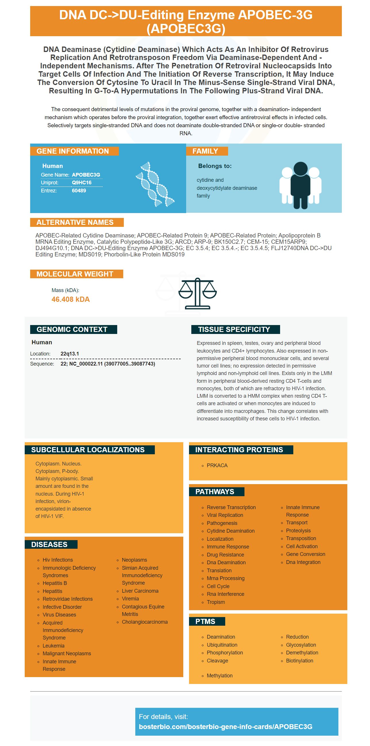
Facts about DNA dC->dU-editing enzyme APOBEC-3G.

The consequent detrimental levels of mutations in the proviral genome, together with a deamination- independent mechanism which operates before the proviral integration, together exert effective antiretroviral effects in infected cells. Selectively targets single-stranded DNA and does not deaminate double-stranded DNA or single-or double- stranded RNA.
| Human | |
|---|---|
| Gene Name: | APOBEC3G |
| Uniprot: | Q9HC16 |
| Entrez: | 60489 |

| Belongs to: |
|---|
| cytidine and deoxycytidylate deaminase family |

APOBEC-related cytidine deaminase; APOBEC-related protein 9; APOBEC-related protein; apolipoprotein B mRNA editing enzyme, catalytic polypeptide-like 3G; ARCD; ARP-9; bK150C2.7; CEM-15; CEM15ARP9; dJ494G10.1; DNA dC->dU-editing enzyme APOBEC-3G; EC 3.5.4; EC 3.5.4.-; EC 3.5.4.5; FLJ12740DNA dC->dU editing enzyme; MDS019; phorbolin-like protein MDS019
Mass (kDA):
46.408 kDA

| Human | |
|---|---|
| Location: | 22q13.1 |
| Sequence: | 22; NC_000022.11 (39077005..39087743) |
Expressed in spleen, testes, ovary and peripheral blood leukocytes and CD4+ lymphocytes. Also expressed in non-permissive peripheral blood mononuclear cells, and several tumor cell lines; no expression detected in permissive lymphoid and non-lymphoid cell lines. Exists only in the LMM form in peripheral blood-derived resting CD4 T-cells and monocytes, both of which are refractory to HIV-1 infection. LMM is converted to a HMM complex when resting CD4 T-cells are activated or when monocytes are induced to differentiate into macrophages. This change correlates with increased susceptibility of these cells to HIV-1 infection.
Cytoplasm. Nucleus. Cytoplasm, P-body. Mainly cytoplasmic. Small amount are found in the nucleus. During HIV-1 infection, virion-encapsidated in absence of HIV-1 VIF.


This article will discuss how scientists can benefit from Steven Boster's research concerning the APOBEC3G gene marker. Learn how to use high-affinity prima antibodies as well as Gene infographics. These are only a few of the many benefits Boster Bio will bring. You can learn more about Steven Boster and his research by clicking the links below.
The APOBEC3G marker can be used to determine the expression levels of a specific protein in human blood. This marker can be used by scientists for specific samples and applications. Scientists can present their findings and receive product credits. Scientists across the globe can apply the Boster method. Scientists can submit results for different species and for applications. The most beneficial uses for markers APOBEC3G do not have to be limited to human patients.
The gene APOBEC3G encodes a protein that has anti-HIV activity. It is a crucial anti-HIV cellular antibody, however, it isn't clear how it is controlled inside the CNS. Therefore, it is important to understand the cellular defense mechanisms as soon as it is possible. The APOBEC3G genes could be another of these factors. A preliminary study has shown that APOBEC3G could be a novel antiviral restriction gene.
Researchers have discovered a brand new mutational signature for APOBEC3G. Previous mutational signatures included W1 or W2 or 13. These signatures have proven to be specific for patients suffering from DCB. The mutational signature W3 has the same APOBEC type that is found in the APOBEC genes. It has been linked to smoking which could explain why it is present in DCB patients.
APOBEC3G is a member of the family of cellular deaminases of cytidine. Its antiretroviral activity requires packaging into virions, and ensuring close association with the nascent retroviral cDNA. APOBEC3G has direct interactions with the gag polyprotein of HIV-1. This interaction is crucial to encapsidation. The N-terminal region (Gag) is the most important.
The clinical outcomes of ovarian cancers and cervical cancers are associated with APOBEC3G gene expression. Furthermore, two studies have revealed that APOBEC3G is associated with an immune-therapy response to cancer. However, more research is needed to fully determine the function of this gene in the development of cancer. We have discovered several gene expression signatures that are highly correlated with clinical outcomes.
In recent years, a number of laboratories have developed high-affinity primary antibodies against APOBEC3G by using EMSA. This combination of electrophoresis and image scanning is called EMSA. The input to EMSA must be controlled, and binding isotherms are determined in relation to a fixed input nucleic acid. High-affinity primary antibodies using the APOBEC3G marker provide many advantages.
The high-affinity primary antibodies eecioy the APOBEC3G marker as a molecular weight marker. Their affinity is high, which boosts the affinity of the antibody and improves the staining performance. To create a variety of dilutions, the antibodies were dispersed at 5 to 25 ug/mL. These antibodies should be tested with samples stained with different amounts and times to ensure they can penetrate the tissue.
The APOBEC3G marker has both anti and pro-HIV actions. The A3G is bound to the viral genome's DNA minus-strand. The virus is represented by green circles in T cells that are infected and red circles outside. It has two copies (RNA genome) and multiple copies (A3G). Cytotoxic T cells with CD8+ recognize this epitope in the context of the MHC class I antigen.
There are many commercial sources for these high-affinity primary antibodies. The quality of the antibody will determine the success or failure of the experiment. You can independently test the quality of the antibody with the literature or by conducting tests. You can also purchase directly from the commercial source. This ensures the quality control of the antibody. If the manufacturer does not provide this information, the data can easily be viewed.
After identifying the lane of interest in the APOBEC3G-labeled Gel, you should cut the strips off. Then, transfer them to the weight boat made of plastic. Then, you should incubate the gel strip with 30 mM DTBP, dimethyl 3,3'-dithiobispropionamide, supplied by Thermo Fisher Scientific. It is also possible to use the TEA-denature method to determine size of the protein complexes that are cross-linked.
In addition to KD values The antibodies are also identified by their affinity factor which explains how fast they connect to their targets. The affinity factor (KD) of the antibody is the ratio of the off-rate and the on-rate of the solution of peptide. Once the target antibody binds to the peptide, the antigen is freed. High-affinity primary antibodies can be utilized for research.
Human genomes include the gene APOBEC3G. The gene is available in the UCSC Genome Browser. To view annotations and other information, click on a grid cell. The informationgraphic of the APOBEC3G gene can be an excellent resource for finding out more about this marker. It is unique in its design. Genes are expressed in many ways and are present throughout the human body.
PMID: 14557625 by Kao S., et al. The human immunodeficiency virus type 1 Vif protein reduces intracellular expression and inhibits packaging of APOBEC3G (CEM15), a cellular inhibitor of virus infectivity.
PMID: 11863358 by Jarmuz A., et al. An anthropoid-specific locus of orphan C to U RNA-editing enzymes on chromosome 22.