This website uses cookies to ensure you get the best experience on our website.
- Table of Contents
6 Citations 19 Q&As
6 Citations 16 Q&As


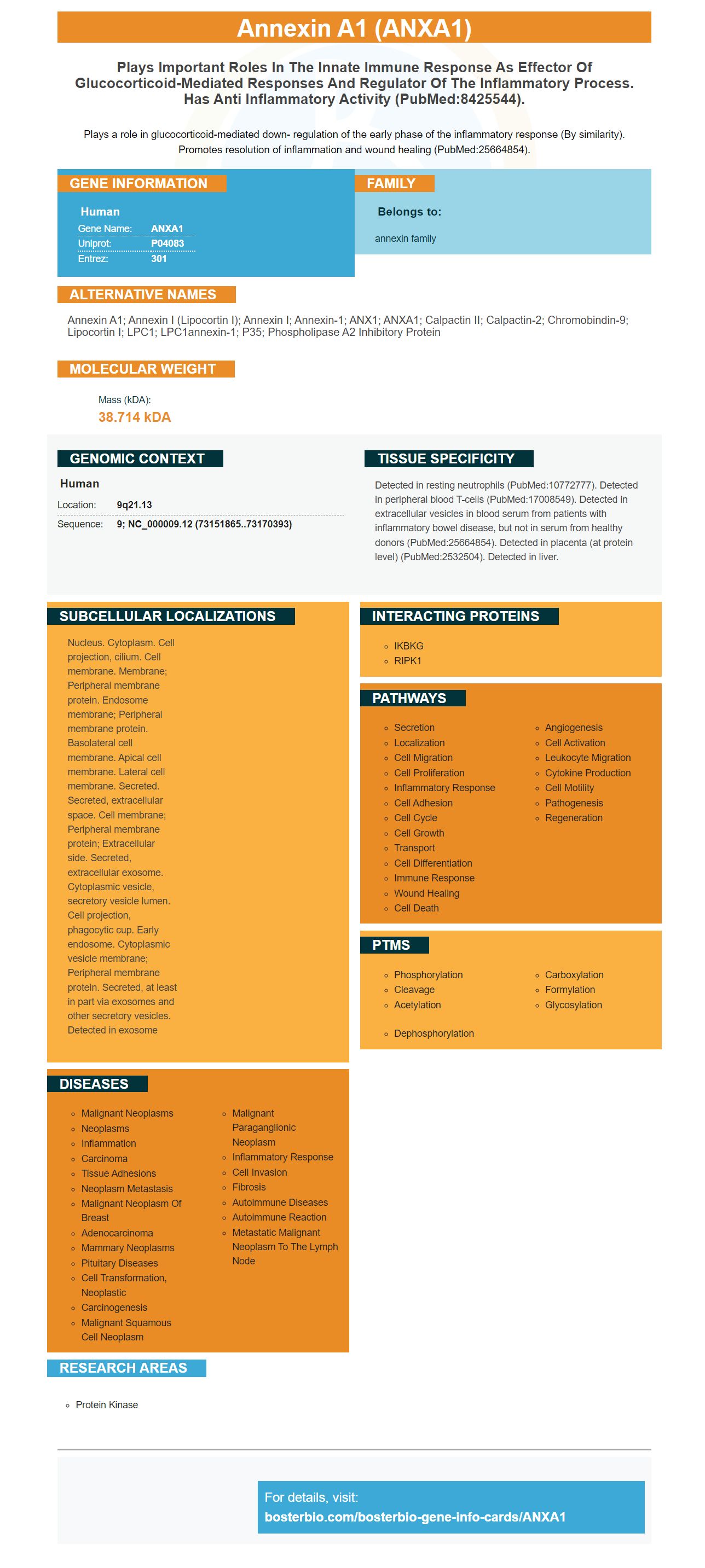
Facts about Annexin A1.

Plays a role in glucocorticoid-mediated down- regulation of the early phase of the inflammatory response (By similarity). Promotes resolution of inflammation and wound healing (PubMed:25664854).
| Human | |
|---|---|
| Gene Name: | ANXA1 |
| Uniprot: | P04083 |
| Entrez: | 301 |

| Belongs to: |
|---|
| annexin family |

Annexin A1; annexin I (lipocortin I); Annexin I; Annexin-1; ANX1; ANXA1; Calpactin II; calpactin-2; chromobindin-9; Lipocortin I; LPC1; LPC1annexin-1; p35; Phospholipase A2 inhibitory protein
Mass (kDA):
38.714 kDA

| Human | |
|---|---|
| Location: | 9q21.13 |
| Sequence: | 9; NC_000009.12 (73151865..73170393) |
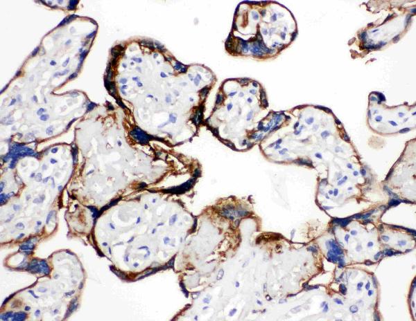
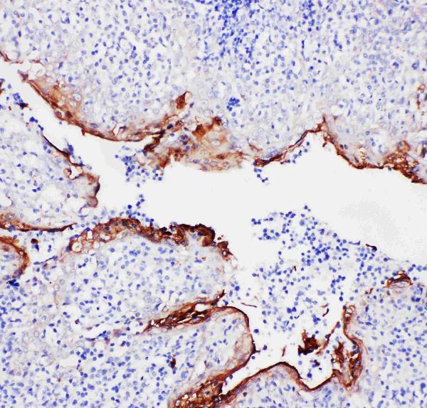
Detected in resting neutrophils (PubMed:10772777). Detected in peripheral blood T-cells (PubMed:17008549). Detected in extracellular vesicles in blood serum from patients with inflammatory bowel disease, but not in serum from healthy donors (PubMed:25664854). Detected in placenta (at protein level) (PubMed:2532504). Detected in liver.
Nucleus. Cytoplasm. Cell projection, cilium. Cell membrane. Membrane; Peripheral membrane protein. Endosome membrane; Peripheral membrane protein. Basolateral cell membrane. Apical cell membrane. Lateral cell membrane. Secreted. Secreted, extracellular space. Cell membrane; Peripheral membrane protein; Extracellular side. Secreted, extracellular exosome. Cytoplasmic vesicle, secretory vesicle lumen. Cell projection, phagocytic cup. Early endosome. Cytoplasmic vesicle membrane; Peripheral membrane protein. Secreted, at least in part via exosomes and other secretory vesicles. Detected in exosome


If you are looking to determine the most effective use for the ANXA1 Marker, you may be wondering how to use it effectively. ANXA1 is a chemical that regulates cell division proliferative, apoptosis, and cell growth. This marker has great potential as a therapeutic target for aggressive cancers. In the end, this antibody has been extensively studied and is now a highly popular and promising biomarker.
ANXA1 is a cytoskeletal proteins that regulates cell proliferation as well as the process of apoptosis. It is involved in the recruitment of monocytes to the site of inflammation, and is essential for orderly progress of acute inflammation to its resolution. AnxA1's Efferocytotic function facilitates the clearing of effete neutrophils at the site of injury. It also prevents macrophages from releasing proinflammatory mediators, and also reduces the tumor-promoting IL 23.
The expression of ANXA1 was examined in gastrointestinal cancers of the human body and non-cancerous tissue. The bile drain was used as a calibrator. After normalizing the mRNA levels fold changes were calculated by divising the ratio of tumor cells to healthy tissue. Western blotting was employed to determine the immunoreactivity of ANXA1 using antibodies against GES1 and anti-actin.
Despite its wide-ranging role in human disease, the exact function of ANXA1 isn't completely understood. Despite the fact that ANXA1 is a regulator of cell growth, a recent study discovered that it blocks breast cancer's EMT pathway. The knockdown of ANXA1 in murine mammary epithelial cells increased the growth of tumors. The ANXA1 gene also suppressed autophagy. This promoted tumor growth as well as lung metastases.
ANXA1 is a gene that is expressed by cancer cells. A lentivirus containing a short-hairpin RNA that targets ANXA1 was used in an experiment to transfect BLCA cells. The cells were then treated with puromycin, and exposed to the lentivirus sh-ANXA1. The results revealed that the expression of ANXA1 had been strongly related to survival.
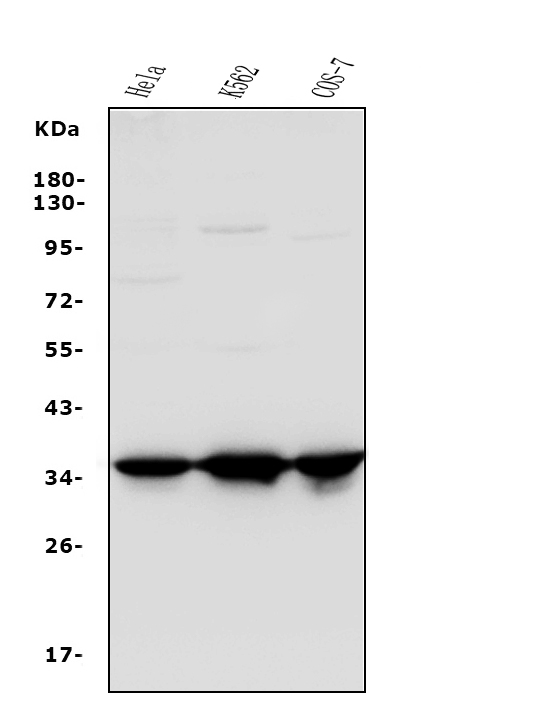
Antibodies to AnnexinA1 are extremely sensitive. Boster Bio Anti-Annexin A1 antibodies can be used to detect this protein in ICC, ELISA and WB applications. They are sensitive and detect levels as low as one picogram or milligram. They can also be used for measuring the expression of other proteins in tumor cells.
Previous studies have revealed that ANXA1 expression in BLCA cells is associated with survival in tumors and poor differentiation, as well as an elevated T status, and increased T status. In a follow-up study, the molecule inhibited tumor growth in xenografts and decreased the survival of patients with no metastasis. The knockdown of ANXA1 led to the expression of EGFR and accelerated ubiquitination of P-EGFR, a known tumor-suppressing protein.
Researchers from Mirimus Inc. in Brooklyn, New York, say a protein known as TMEM230 could be a therapeutic target for cancers that are aggressive. In particular this protein could be effective against glioblastoma, the most deadly form of brain cancer. A majority of these tumors are not viable therapeutic agents because they have blood vessels that are abnormal which makes them difficult to focus on. The discovery of a novel target is crucial for the successful treatment of tumors that are aggressive. Endogenous gene expression inhibitors might be less toxic than small-molecule medications and less likely to cause resistance in tumors.
Although little research has been done to identify predictive biomarkers in TNBC (or any other type of cancer) research suggests that the microRNA-regulated transcriptional dynamics plays a significant role in metastasis. MicroRNA-based treatments for cancer target a variety of pathways. Researchers hope to improve the outcome for patients with aggressive cancers by targeting microRNAs.
This Boster Bio Anti-Annexin-A1 Pathology antibody was tested in IHC, ICC, and WB applications. The antibody reacts with Human Annexin A1. This protein is involved in innate and adaptive immune system and is a crucial player in the biosynthesis of mediators, prostaglandins and inflammation. It is also absent in some B-cell lymphomas.
Boster provides high-quality monoclonal antibodies that have been validated to a variety of purposes. They are designed in-house, and sourced from well-known clones. The antibodies can also be conjugated to IHC/IF, flow Cytometry and ChIP. Once you've purchased your Boster Bio ANXA1 Marker, you'll need to select the conjugate during the checkout procedure.
The results of this study suggest that ANXA1 is an excellent biomarker for BLCA. It stimulates EGFR signaling and promotes growth and migration. More research is needed to determine if this marker could be used as a biomarker to assist patients suffering from this disease. If this is successful, it could lead to a personalised treatment for BLCA.
When it comes down to making high-quality antibodies and ELISA kits, Boster Bio excels. Boster Bio's lab has specialized capabilities to provide well-tested reagents, antibodies, and other products to the research community. Boster Bio's products have over 29,000 citations and are designed for WB and ELISA use. Boster Bio offers antibodies and ELISA kit that can be used to research in the fields of neuroscience, cancer research, and other fields. In addition, Boster offers a Quality Guarantee, which guarantees that each product will deliver as promised.
The Boster bio product line includes reagents as well as lysates. Boster also offers a variety of kits for assays that are popular. Many of these reagents are validated by WB so you can be confident that they'll work in your test. Many of these products can be customized to meet your exact requirements. They also offer technical support to make sure you get the most benefit from your experiments.
Founded in 1993, Boster Bio is a manufacturer of specific and high-sensitivity antibodies. The company has spent the last two years perfecting its technology and methods. Over 20,000 publications have featured Boster Bio's products. Every Boster Bio product is validated on WB, ELISA, Flow Cytometry, and IHC. Furthermore Boster Bio's antibody are tested against 250 human and animal tissues and are therefore remarkably sensitive and specific.
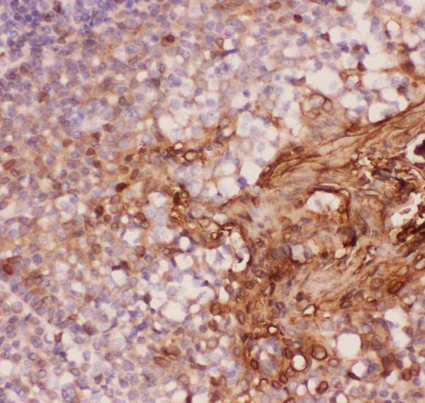
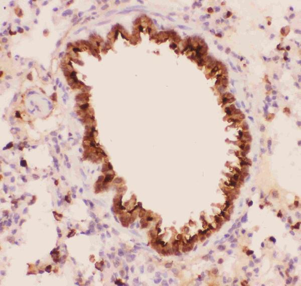
The ANXA1 marker is used to perform immunohistochemistry (IHC). The process involves placing a sample in a neutral gel, and then cutting it using a rotary microtome, and staining it. The antigen is visualized within the section. The tissue sample should be dehydrated to ensure long-term storage. There could be variations in the chromogens used, counterstains, enzymes, and glycerol that are used to stain IHC stains.
Many biological activities are associated with the ANXA1 gene. It is anti-inflammatory and has proliferative properties and regulates cell movement and is associated to tumorigenesis. In addition to its prognostic significance, ANXA1 is also implicated in the carcinogenesis process. It has not been shown to be a reliable method to predict the size of tumors, however, it does have prognostic value.
ANXA1 expression was studied in six human malignancies. ANXA1 expression was also lower in esophageal and colon cancers. It was significantly higher in pancreatic cancers as well as esophageal carcinomas. In addition pancreatic cancers showed a 1.6-fold increase in mRNA levels as well as ANXA1 protein expression when compared to the surrounding tissue. However, prostate and colorectal cancer showed no significant changes in ANXA1 expression.
The judge evaluates the state's capacity and willingness to conduct lawful proceedings when the case is brought to the ICC. This is done by looking at evidence from the state as well as national investigations. If the state was unwilling or unable to prosecute, it's considered admissible. ICC judges decide if the case is similar as the prior one. To be able to validate the case, it must be identical to the preceding one.
ICC is an index used to measure the level of agreement between different measurements. It is extensively used in traditional medicine. It is a test for tests-retests and intrarater reliability. There are ten different forms of ICC that each have different assumptions and interpretations. Researchers should understand the principles of deciding on the correct one. They should present the true ICC instead of an estimate. Furthermore, ICC should be reported as 95% confidence interval.
The cells are stained using the specific antibody and nuclear probe DAPI for this test. Then, the cells are examined using a confocal camera. The images gathered using this method are extremely detailed and contain high-resolution three-color resolution. For each antibody that has been ICC-validated, multiple multicolor images are included in the product data pages. They show the target protein's expression within different subcellular regions.
PMID: 2936963 by Wallner B.P., et al. Cloning and expression of human lipocortin, a phospholipase A2 inhibitor with potential anti-inflammatory activity.
PMID: 1832554 by Kovacic R.T., et al. Correlation of gene and protein structure of rat and human lipocortin I.
*More publications can be found for each product on its corresponding product page