This website uses cookies to ensure you get the best experience on our website.
- Table of Contents
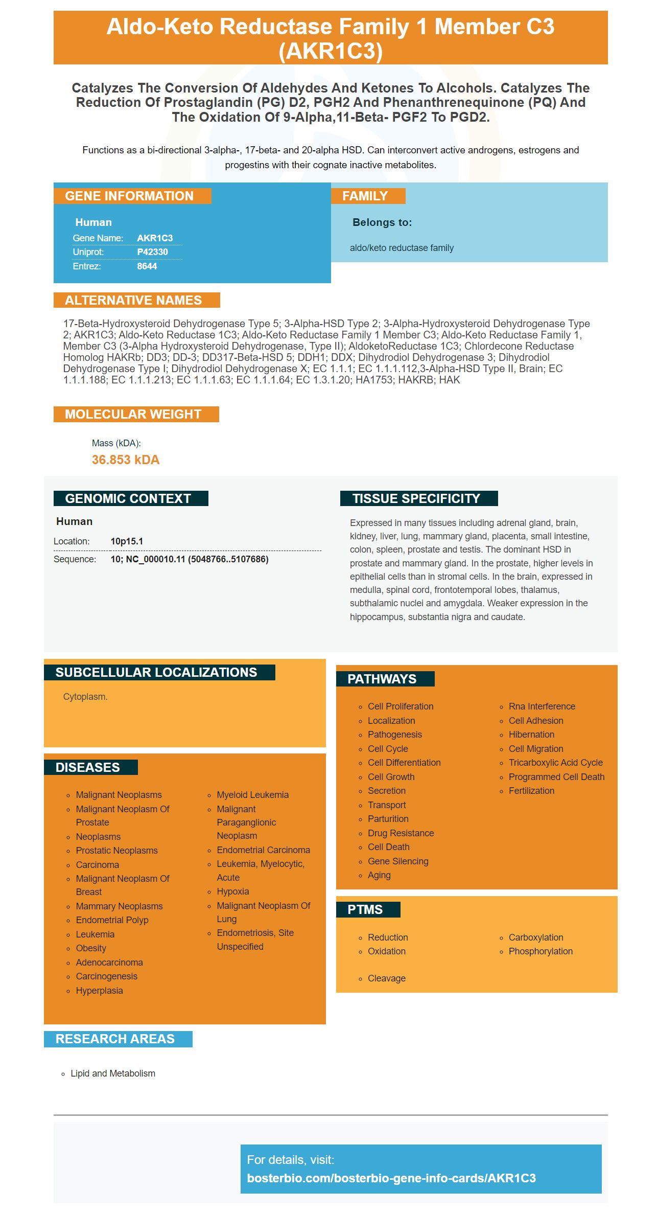


Facts about Aldo-keto reductase family 1 member C3.

Functions as a bi-directional 3-alpha-, 17-beta- and 20-alpha HSD. Can interconvert active androgens, estrogens and progestins with their cognate inactive metabolites.
| Human | |
|---|---|
| Gene Name: | AKR1C3 |
| Uniprot: | P42330 |
| Entrez: | 8644 |

| Belongs to: |
|---|
| aldo/keto reductase family |

17-beta-hydroxysteroid dehydrogenase type 5; 3-alpha-HSD type 2; 3-alpha-hydroxysteroid dehydrogenase type 2; AKR1C3; Aldo-keto Reductase 1C3; aldo-keto reductase family 1 member C3; aldo-keto reductase family 1, member C3 (3-alpha hydroxysteroid dehydrogenase, type II); AldoketoReductase 1C3; Chlordecone reductase homolog HAKRb; DD3; DD-3; DD317-beta-HSD 5; DDH1; DDX; Dihydrodiol dehydrogenase 3; Dihydrodiol dehydrogenase type I; dihydrodiol dehydrogenase X; EC 1.1.1; EC 1.1.1.112,3-alpha-HSD type II, brain; EC 1.1.1.188; EC 1.1.1.213; EC 1.1.1.63; EC 1.1.1.64; EC 1.3.1.20; HA1753; HAKRB; HAK
Mass (kDA):
36.853 kDA

| Human | |
|---|---|
| Location: | 10p15.1 |
| Sequence: | 10; NC_000010.11 (5048766..5107686) |
Expressed in many tissues including adrenal gland, brain, kidney, liver, lung, mammary gland, placenta, small intestine, colon, spleen, prostate and testis. The dominant HSD in prostate and mammary gland. In the prostate, higher levels in epithelial cells than in stromal cells. In the brain, expressed in medulla, spinal cord, frontotemporal lobes, thalamus, subthalamic nuclei and amygdala. Weaker expression in the hippocampus, substantia nigra and caudate.
Cytoplasm.


The AKR1C3 gene is an important target in many different hormone-dependent diseases. Moreover, it is also involved in the regulation of PDGF-A, NRF2, and CYP17A1 in the human body. To use this gene in research and development, you should read this article. It is applicable to all scientists worldwide. Read on to learn more about its best uses.
The AKR1C3 gene is a major epigenetic regulator in prostate cancer. AKR1C3-selective inhibitors can be used in combination with HDAC inhibition. Standard treatment for LARC patients includes preoperative chemoradiotherapy. While preoperative chemoradiotherapy results in a complete pathological response, approximately 20% to 40% of patients have a partial or absent response to treatment.
The gene encoding AKR1C3 is found in multiple tissues, including the testes. It is expressed in the common ancestor of both animals and fungi. In addition to being expressed in the testes, it also is found in the extragonadal tissues of humans. It catalyses the NADPH-dependent reduction of AD to T. Moreover, it is involved in the syntheses of two prostaglandins, T and DHT.
The sulphonamide ring is found to be in close proximity to a structured water molecule in the SP3 pocket. By studying morpholino(phenylpiperazin-2-one) derivatives with different N-substituted triazoles, Heinrich et al., identified a new inhibitor of AKR1C3 that inhibits the enzyme without interacting with the oxyanion hole. This compound was found to be significantly more active than its carboxylic acid analogue 18 and exhibited low off-target inhibition of COX1 and COX2.
Inhibitors that are more selective against AKR1C3 have been identified through molecular docking. The activity of the compounds identified in these studies correlates with their inhibitory activity. However, there is a need for more drug-like compounds that inhibit AKR1C3 in humans. If these compounds are developed, they may be used in clinical trials.
Researchers hope that selective inhibitors of HSD17B3 will be more effective than existing endocrine therapies in PCa. In addition, the use of selective HSD17B3 inhibitors could reduce off-target effects in patients with PCa. The combination of HSD17B3 inhibitors with AKR1C3 selective inhibitors may result in an enhanced inhibition of the biosynthetic pathway and AR binding in PCa patients.
Despite being implicated in several cancers, AKR1C3 is especially prevalent in prostate malignancy. Prostate cancer cells express AKR1C3 proteins, which contribute to androgen metabolism and downstream stimulation of the androgen receptor. Although cancer cells have different targets, they all show elevated expression levels of AKR1C3 in different tissues.
PDGF-A is a ligand for the receptor AKR1C3, which is expressed by various asthma pathogenesis cells. It is involved in the regulation of inflammatory response and airway remodeling, leading to a range of symptoms. Various proteins in the platelet-derived growth factor family bind to PDGF receptors and activate signal transducers. Akt is one such transcription factor.
AKR1C3 is a member of the aldo-keto reductase family and reduces carbonyl substrates, including keto-steroids, fatty acids, and lipid peroxidation byproducts. The enzyme also reduces retinals and quinones. This enzyme may play a role in preventing OS cell proliferation.
The PDGF receptor family consists of heterodimers and homodimers, which are all derived from the same precursor molecule. PDGF receptors are structurally similar, with extracellular domains with five immunoglobulin-like domains and intracellular parts with kinase domains. These are homodimers of the receptor, with domains 2 and 3 binding ligands. The remaining domains form a dimer and are autophosphorylated by the PDGFRa and PDGF-B.
PDGF receptors are implicated in the promotion of tumorigenesis and angiogenesis in many types of cancer. In particular, stromal PDGF receptors have been associated with poor prognosis in breast, pancreatic, and colorectal cancer. Moreover, tumors containing these receptors show increased growth and metastasis, and the expression of AKR1C3 in these cancers is associated with poorer outcomes.
T-cell proliferation is negatively regulated by Cbl, a protein with a tyrosine kinase domain. This protein also negatively regulates platelet derived growth factor receptor-dependent cell proliferation. Wang Y and Wang Z have investigated the role of Cbl in platelet derived growth factor receptor signaling. Their recent study suggests that a similar role for AKR1C3 in platelet stem cell proliferation.
The PDGF receptor a and b are expressed in tumors and stromal cells. These two proteins are required for neural crest cell development and somite patterning. PDGF-A has also been implicated in lung alveogenesis, as demonstrated in Bostrom and Karlsson. In addition, AKR1C3 amplification has been observed in a range of cancer types, including glioblastoma multiforme, oligodendrogliomas, and squamous cell carcinoma.
The AKR1C3 marker has a very specific use in cytochemistry, as it is involved in the regulation of cell growth and division. However, it is not completely clear how this marker contributes to these processes. In fact, it may not even be necessary to test for this protein in vivo. Boster offers a series of high-affinity primary antibodies, including the AKR1C3 marker, that have been widely cited in the scientific community. Boster's antibodies are validated by ELISA and Western Blotting, so you can be confident in the quality of your results.
CRPC patients are at high risk for AKR1C3 levels. The protein may also be used as a biomarker for intratumoral steroidogenesis. AKR1C3 is found in biopsy and transurethral resection specimens. DuCaP is a protein that converts DHEA into T and DHT. Fortunately, it is possible to inhibit AKR1C3 activity by using drugs that block the activity of this enzyme.
The aldo-keto reductase B10 protein has been cloned, expressed and purified. It is related to aflatoxin dialdehyde reductases and has been placed in a novel family of enzymes known as AKR14. The sequence of this enzyme is found in many other bacteria. It reduces the toxic metabolite methylglyoxal.
The AKR1C3 protein is a pre-receptor regulator of membrane and nuclear receptors. Changing the levels of this protein is related to the development of endometrial cancer, prostate cancer, and other diseases. In addition, mutations in AKR1C1 and AKR1D1 cause sexual development dysgenesis. It is also a causative factor of bile-acid deficiency.
The steroidogenic pathway requires the enzyme CYP17A1. This P450 is expressed in the adrenal gland and gonads and is controlled by a complex interaction between transcription factors. It is not directly affected by epigenetic regulation, as it lacks CpG islands. However, the presence of 5aza-dC in the human CYP17A1 gene suggests indirect epigenetic control. CYP17A1 has two activities: lyase and 17a-hydroxylase.
The ring of morpholine is located within hydrogen bonding distance of a structured water molecule that is part of the SP3 pocket. The importance of this oxygen was studied by examining derivatives of morpholine ring bound to urea. The compounds that did not contain morpholine ring were inactive. Meanwhile, those with larger substituents exhibited higher activity than their inactive counterparts.
Mutant AKR1C3 partially rescues growth and colony formation in Siah2 KD Rv1 cells. In addition, AKR1C3 re-expression partially restored colony formation in Siah2 KD Rv1 cells. Thus, AKR1C3 has a role in the regulation of Siah2-dependent cell proliferation.
While the function of AKR1C3 in regulating androgen levels is unknown, it has been identified in normal prostate cells. Its significance seems to be related to its ability to reduce prostaglandin D2 to F2 and synthesis of T. The activity of CYP17A1 is essential for the metabolism of various hormones in the body.
A better understanding of the PCa microenvironment will help improve treatment options. Knowing how AKR1C3 is controlled in the tumour microenvironment will improve targeting of key enzymes in the steroidogenic pathway. Its role in CRPC is uncertain. If identified, AKR1C3 may be an effective therapeutic target. In addition, its effect on prostate cancer cells has yet to be determined.
In addition to AKR1C3, HSD17B3 belongs to the SDR family and catalyzes the same reaction as AKR1C3. The crystal structure of AKR1C3 was determined by Lovering et al. It revealed a typical aldo-keto reductase structure, as well as a steroid channel SC.
PMID: 8274401 by Qin K.-N., et al. Molecular cloning of multiple cDNAs encoding human enzymes structurally related to 3 alpha-hydroxysteroid dehydrogenase.
PMID: 7650035 by Khanna M., et al. Substrate specificity, gene structure, and tissue-specific distribution of multiple human 3 alpha-hydroxysteroid dehydrogenases.


