Click image to see more details
Product Info Summary
| SKU: | PA1639 |
|---|---|
| Size: | 100 μg/vial |
| Reactive Species: | Human, Mouse, Rat |
| Host: | Rabbit |
| Application: | IHC, ICC, WB |
Customers Who Bought This Also Bought

Product info
Product Name
Anti-XRCC1 Antibody
SKU/Catalog Number
PA1639
Size
100 μg/vial
Form
Lyophilized
Description
Boster Bio Anti-XRCC1 Antibody catalog # PA1639. Tested in IHC, ICC, WB applications. This antibody reacts with Human, Mouse, Rat.
Storage & Handling
Store at -20˚C for one year from date of receipt. After reconstitution, at 4˚C for one month. It can also be aliquotted and stored frozen at -20˚C for six months. Avoid repeated freeze-thaw cycles.
Cite This Product
Anti-XRCC1 Antibody (Boster Biological Technology, Pleasanton CA, USA, Catalog # PA1639)
Host
Rabbit
Contents
Each vial contains 5mg BSA, 0.9mg NaCl, 0.2mg Na2HPO4, 0.05mg Thimerosal, 0.05mg NaN3.
Clonality
Polyclonal
Isotype
Rabbit IgG
Immunogen
A synthetic peptide corresponding to a sequence at the N-terminus of human XRCC1, identical to the related mouse sequence, and different from the related rat sequence by one amino acid.
*Blocking peptide can be purchased. Costs vary based on immunogen length. Contact us for pricing.
Cross-reactivity
No cross-reactivity with other proteins
Reactive Species
PA1639 is reactive to XRCC1 in Human, Mouse, Rat
Applications
PA1639 is guaranteed for IHC, ICC, WB Boster Guarantee
Observed Molecular Weight
90 kDa
Calculated molecular weight
69477 MW
Background of XRCC1
XRCC1 (X-RAY REPAIR, COMPLEMENTING DEFECTIVE, IN CHINESE HAMSTER, 1) is a DNA repair protein which complexes with DNA ligase III. The protein encoded by this gene is involved in the efficient repair of DNA single-strand breaks formed by exposure to ionizing radiation and alkylating agents. The XRCC1 gene is mapped to 19q13.31. The XRCC1 interacts with DNA ligase III, polymerase beta and poly (ADP-ribose) polymerase to participate in the base excision repair pathway. It may play a role in DNA processing during meiogenesis and recombination in germ cells. A rare microsatellite polymorphism in this gene is associated with cancer in patients of varying radiosensitivity. XRCC1 is phosphorylated in vivo and in vitro by CK2, and CK2 phosphorylation of XRCC1 on ser518, thr519, and thr523 largely determines aprataxin binding to XRCC1 through its FHA domain.
Antibody Validation
Boster validates all antibodies on WB, IHC, ICC, Immunofluorescence, and ELISA with known positive control and negative samples to ensure specificity and high affinity, including thorough antibody incubations.
Assay dilution & Images
Reconstitution
Add 0.2ml of distilled water will yield a concentration of 500ug/ml.
Assay Dilutions Recommendation
The recommendations below provide a starting point for assay optimization. The actual working concentration varies and should be decided by the user.
Immunocytochemistry , 0.5-1μg/ml, Human, Mouse, Rat
Immunohistochemistry (Paraffin-embedded Section), 0.5-1μg/ml, Human, Mouse, Rat, By Heat
Western blot, 0.1-0.5μg/ml, Human, Mouse, Rat
Validation Images & Assay Conditions
Click image to see more details
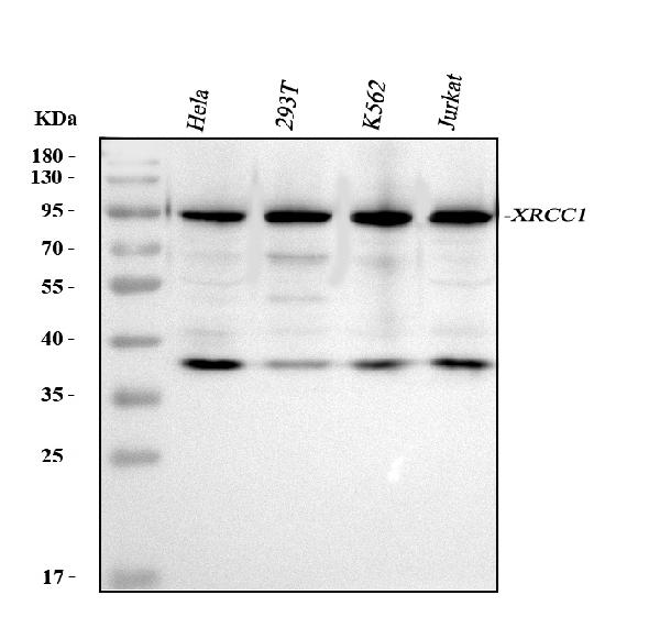
Figure 1. Western blot analysis of XRCC1 using anti-XRCC1 antibody (PA1639).
Electrophoresis was performed on a 5-20% SDS-PAGE gel at 70V (Stacking gel) / 90V (Resolving gel) for 2-3 hours. The sample well of each lane was loaded with 30 ug of sample under reducing conditions.
Lane 1: human Hela whole cell lysates,
Lane 2: human 293T whole cell lysates,
Lane 3: human K562 whole cell lysates,
Lane 4: human Jurkat whole cell lysates.
After electrophoresis, proteins were transferred to a nitrocellulose membrane at 150 mA for 50-90 minutes. Blocked the membrane with 5% non-fat milk/TBS for 1.5 hour at RT. The membrane was incubated with rabbit anti-XRCC1 antigen affinity purified polyclonal antibody (Catalog # PA1639) at 0.5 μg/mL overnight at 4°C, then washed with TBS-0.1%Tween 3 times with 5 minutes each and probed with a goat anti-rabbit IgG-HRP secondary antibody at a dilution of 1:5000 for 1.5 hour at RT. The signal is developed using an Enhanced Chemiluminescent detection (ECL) kit (Catalog # EK1002) with Tanon 5200 system. A specific band was detected for XRCC1 at approximately 90 kDa. The expected band size for XRCC1 is at 90 kDa.

Click image to see more details
Figure 2. IHC analysis of XRCC1 using anti-XRCC1 antibody (PA1639).
XRCC1 was detected in a paraffin-embedded section of Human Mammary Cancer tissue. Heat mediated antigen retrieval was performed in EDTA buffer (pH 8.0, epitope retrieval solution). The tissue section was blocked with 10% goat serum. The tissue section was then incubated with 1 μg/ml rabbit anti-XRCC1 Antibody (PA1639) overnight at 4°C. Peroxidase Conjugated Goat Anti-rabbit IgG was used as secondary antibody and incubated for 30 minutes at 37°C. The tissue section was developed using HRP Conjugated Rabbit IgG Super Vision Assay Kit (Catalog # SV0002) with DAB as the chromogen.

Click image to see more details
Figure 3. ICC analysis of XRCC1 using anti-XRCC1 antibody (PA1639).
XRCC1 was detected in an immunocytochemical section of A549 cells. Enzyme antigen retrieval was performed using IHC enzyme antigen retrieval reagent (AR0022) for 15 mins. The cells were blocked with 10% goat serum. And then incubated with 1 μg/ml rabbit anti-XRCC1 Antibody (PA1639) overnight at 4°C. Biotinylated goat anti-rabbit IgG was used as secondary antibody and incubated for 30 minutes at 37°C. The section was developed using Strepavidin-Biotin-Complex (SABC)(Catalog # SA1022) with DAB as the chromogen.
Protein Target Info & Infographic
Gene/Protein Information For XRCC1 (Source: Uniprot.org, NCBI)
Gene Name
XRCC1
Full Name
DNA repair protein XRCC1
Weight
69477 MW
Alternative Names
DNA repair protein XRCC1; RCC; X-ray repair complementing defective repair in Chinese hamster cells 1; X-ray repair cross-complementing protein 1; X-ray-repair, complementing defective, repair in Chinese hamster XRCC1 RCC, SCAR26 X-ray repair cross complementing 1 DNA repair protein XRCC1|X-ray repair complementing defective repair in Chinese hamster cells 1|X-ray repair cross-complementing protein 1
*If product is indicated to react with multiple species, protein info is based on the gene entry specified above in "Species".For more info on XRCC1, check out the XRCC1 Infographic

We have 30,000+ of these available, one for each gene! Check them out.
In this infographic, you will see the following information for XRCC1: database IDs, superfamily, protein function, synonyms, molecular weight, chromosomal locations, tissues of expression, subcellular locations, post-translational modifications, and related diseases, research areas & pathways. If you want to see more information included, or would like to contribute to it and be acknowledged, please contact [email protected].
Specific Publications For Anti-XRCC1 Antibody (PA1639)
Hello CJ!
No publications found for PA1639
*Do you have publications using this product? Share with us and receive a reward. Ask us for more details.
Recommended Resources
Here are featured tools and databases that you might find useful.
- Boster's Pathways Library
- Protein Databases
- Bioscience Research Protocol Resources
- Data Processing & Analysis Software
- Photo Editing Software
- Scientific Literature Resources
- Research Paper Management Tools
- Molecular Biology Software
- Primer Design Tools
- Bioinformatics Tools
- Phylogenetic Tree Analysis
Customer Reviews
Have you used Anti-XRCC1 Antibody?
Submit a review and receive an Amazon gift card.
- $30 for a review with an image
0 Reviews For Anti-XRCC1 Antibody
Customer Q&As
Have a question?
Find answers in Q&As, reviews.
Can't find your answer?
Submit your question
17 Customer Q&As for Anti-XRCC1 Antibody
Question
We bought anti-XRCC1 antibody for WB on eye a few months ago. I am using human, and We intend to use the antibody for IHC next. We are interested in examining eye as well as cervix carcinoma in our next experiment. Could you please give me some suggestion on which antibody would work the best for IHC?
Verified Customer
Verified customer
Asked: 2020-04-10
Answer
I took a look at the website and datasheets of our anti-XRCC1 antibody and it appears that PA1639 has been validated on human in both WB and IHC. Thus PA1639 should work for your application. Our Boster satisfaction guarantee will cover this product for IHC in human even if the specific tissue type has not been validated. We do have a comprehensive range of products for IHC detection and you can check out our website bosterbio.com to find out more information about them.
Boster Scientific Support
Answered: 2020-04-10
Question
Is this PA1639 anti-XRCC1 antibody reactive to the isotypes of XRCC1?
Verified Customer
Verified customer
Asked: 2020-03-30
Answer
The immunogen of PA1639 anti-XRCC1 antibody is A synthetic peptide corresponding to a sequence at the N-terminus of human XRCC1(15-34aa QDSTHCAENLLKADTYRKWR), identical to the related mouse sequence, and different from the related rat sequence by one amino acid. Could you tell me which isotype you are interested in so I can help see if the immunogen is part of this isotype?
Boster Scientific Support
Answered: 2020-03-30
Question
We are currently using anti-XRCC1 antibody PA1639 for human tissue, and we are happy with the IHC results. The species of reactivity given in the datasheet says human, mouse, rat. Is it possible that the antibody can work on zebrafish tissues as well?
Verified Customer
Verified customer
Asked: 2020-02-13
Answer
The anti-XRCC1 antibody (PA1639) has not been validated for cross reactivity specifically with zebrafish tissues, though there is a good chance of cross reactivity. We have an innovator award program that if you test this antibody and show it works in zebrafish you can get your next antibody for free. Please contact me if I can help you with anything.
Boster Scientific Support
Answered: 2020-02-13
Question
We have been able to see staining in rat right ovary. Do you have any suggestions? Is anti-XRCC1 antibody supposed to stain right ovary positively?
Verified Customer
Verified customer
Asked: 2019-09-11
Answer
From what I have seen in literature right ovary does express XRCC1. From what I have seen in Uniprot.org, XRCC1 is expressed in right ovary, eye, cervix carcinoma, leukemic t-cell, cervix carcinoma erythroleukemia, liver, among other tissues. Regarding which tissues have XRCC1 expression, here are a few articles citing expression in various tissues:
Cervix carcinoma, Pubmed ID: 17081983, 18669648, 20068231
Cervix carcinoma, and Erythroleukemia, Pubmed ID: 23186163
Eye, Pubmed ID: 15489334
Leukemic T-cell, Pubmed ID: 19690332
Liver, Pubmed ID: 24275569
Boster Scientific Support
Answered: 2019-09-11
Question
Does PA1639 anti-XRCC1 antibody work on parafin embedded sections? If so, which fixation method do you recommend we use (PFA, paraformaldehyde, other)?
Verified Customer
Verified customer
Asked: 2019-08-07
Answer
As indicated on the product datasheet, PA1639 anti-XRCC1 antibody as been tested on ICC. It is best to use PFA for fixation because it has better tissue penetration ability. PFA needs to be prepared fresh before use. Long term stored PFA turns into formalin, as the PFA molecules congregate and become formalin.
Boster Scientific Support
Answered: 2019-08-07
Question
Would anti-XRCC1 antibody PA1639 work on pig IHC with cervix carcinoma?
Verified Customer
Verified customer
Asked: 2019-06-17
Answer
Our lab technicians have not tested anti-XRCC1 antibody PA1639 on pig. You can run a BLAST between pig and the immunogen sequence of anti-XRCC1 antibody PA1639 to see if they may cross-react. If the sequence homology is close, then you can perform a pilot test. Keep in mind that since we have not validated pig samples, this use of the antibody is not covered by our guarantee. However we have an innovator award program that if you test this antibody and show it works in pig cervix carcinoma in IHC, you can get your next antibody for free.
Boster Scientific Support
Answered: 2019-06-17
Question
I see that the anti-XRCC1 antibody PA1639 works with ICC, what is the protocol used to produce the result images on the product page?
Verified Customer
Verified customer
Asked: 2019-03-11
Answer
You can find protocols for ICC on the "support/technical resources" section of our navigation menu. If you have any further questions, please send an email to [email protected]
Boster Scientific Support
Answered: 2019-03-11
Question
I was wanting to use your anti-XRCC1 antibody for ICC for mouse liver on frozen tissues, but I want to know if it has been validated for this particular application. Has this antibody been validated and is this antibody a good choice for mouse liver identification?
Verified Customer
Verified customer
Asked: 2018-12-25
Answer
As indicated on the product datasheet, PA1639 anti-XRCC1 antibody has been tested for IHC, ICC, WB on human, mouse, rat tissues. We have an innovator award program that if you test this antibody and show it works in mouse liver in IHC-frozen, you can get your next antibody for free.
Boster Scientific Support
Answered: 2018-12-25
Question
My colleagues were satisfied with the WB result of your anti-XRCC1 antibody. However we have observed positive staining in eye nucleus using this antibody. Is that expected? Could you tell me where is XRCC1 supposed to be expressed?
Verified Customer
Verified customer
Asked: 2018-12-10
Answer
According to literature, eye does express XRCC1. Generally XRCC1 expresses in nucleus. Regarding which tissues have XRCC1 expression, here are a few articles citing expression in various tissues:
Cervix carcinoma, Pubmed ID: 17081983, 18669648, 20068231
Cervix carcinoma, and Erythroleukemia, Pubmed ID: 23186163
Eye, Pubmed ID: 15489334
Leukemic T-cell, Pubmed ID: 19690332
Liver, Pubmed ID: 24275569
Boster Scientific Support
Answered: 2018-12-10
Question
Is there a BSA free version of anti-XRCC1 antibody PA1639 available?
Verified Customer
Verified customer
Asked: 2017-12-29
Answer
Thank you for your recent telephone inquiry. I can confirm that some lots of this anti-XRCC1 antibody PA1639 are BSA free. For now, these lots are available and we can make a BSA free formula for you free of charge. It will take 3 extra days to prepare. If you require this antibody BSA free again in future, please do not hesitate to contact me and I will be pleased to check which lots we have in stock that are BSA free.
Boster Scientific Support
Answered: 2017-12-29
Question
I am interested in using your anti-XRCC1 antibody for spinocerebellar ataxia studies. Has this antibody been tested with western blotting on hela cell lysate? We would like to see some validation images before ordering.
Verified Customer
Verified customer
Asked: 2017-09-27
Answer
I appreciate your inquiry. This PA1639 anti-XRCC1 antibody is tested on 293t cell lysate, a431 cell lysate, hela cell lysate, mammary cancer tissue. It is guaranteed to work for IHC, ICC, WB in human, mouse, rat. Our Boster guarantee will cover your intended experiment even if the sample type has not been be directly tested.
Boster Scientific Support
Answered: 2017-09-27
Question
I am interested in to test anti-XRCC1 antibody PA1639 on mouse liver for research purposes, then I may be interested in using anti-XRCC1 antibody PA1639 for diagnostic purposes as well. Is the antibody suitable for diagnostic purposes?
H. Huang
Verified customer
Asked: 2016-12-28
Answer
The products we sell, including anti-XRCC1 antibody PA1639, are only intended for research use. They would not be suitable for use in diagnostic work. If you have the means to develop a product into diagnostic use, and are interested in collaborating with us and develop our product into an IVD product, please contact us for more discussions.
Boster Scientific Support
Answered: 2016-12-28
Question
We appreciate helping with my inquiry over the phone. Here are the WB image, lot number and protocol we used for liver using anti-XRCC1 antibody PA1639. Let me know if you need anything else.
K. Banerjee
Verified customer
Asked: 2016-12-05
Answer
We appreciate the data. You have provided everything we needed. Our lab team are working to resolve your inquiry as quickly as possible, and we appreciate your patience and understanding! Please let me know if there is anything you need in the meantime.
Boster Scientific Support
Answered: 2016-12-05
Question
Is a blocking peptide available for product anti-XRCC1 antibody (PA1639)?
V. Evans
Verified customer
Asked: 2016-05-26
Answer
We do provide the blocking peptide for product anti-XRCC1 antibody (PA1639). If you would like to place an order for it please contact [email protected] and make a special request.
Boster Scientific Support
Answered: 2016-05-26
Question
Does anti-XRCC1 antibody PA1639 work for ICC with liver?
T. Jones
Verified customer
Asked: 2016-03-09
Answer
According to the expression profile of liver, XRCC1 is highly expressed in liver. So, it is likely that anti-XRCC1 antibody PA1639 will work for ICC with liver.
Boster Scientific Support
Answered: 2016-03-09
Question
Can you help my question with product PA1639, anti-XRCC1 antibody. I was wondering if it would be possible to conjugate this antibody with biotin. I would need it to be without BSA or sodium azide. I am planning on using a buffer exchange of sodium azide with PBS only. Would there be problems for me to conjugate the antibody and store it in -20 degrees in small aliquots?
G. Jackson
Verified customer
Asked: 2015-12-04
Answer
We do not advise storing this antibody with PBS buffer only in -20 degrees. If you want to store it in -20 degrees it is best to add some cryoprotectant like glycerol. If you want carrier free PA1639 anti-XRCC1 antibody, we can provide it to you in a special formula with trehalose and/or glycerol. These molecules will not interfere with conjugation chemistry and provide a good level of protection for the antibody from degradation. Please be sure to specify this in your purchase order.
Boster Scientific Support
Answered: 2015-12-04
Question
See attached the WB image, lot number and protocol we used for liver using anti-XRCC1 antibody PA1639. Please let me know if you require anything else.
K. Collins
Verified customer
Asked: 2014-01-09
Answer
Thank you very much for the data. Our lab team are working to resolve this as quickly as possible, and we appreciate your patience and understanding! You have provided everything we needed. Please let me know if there is anything you need in the meantime.
Boster Scientific Support
Answered: 2014-01-09
