This website uses cookies to ensure you get the best experience on our website.
- Table of Contents
1 Citations 16 Q&As
16 Q&As
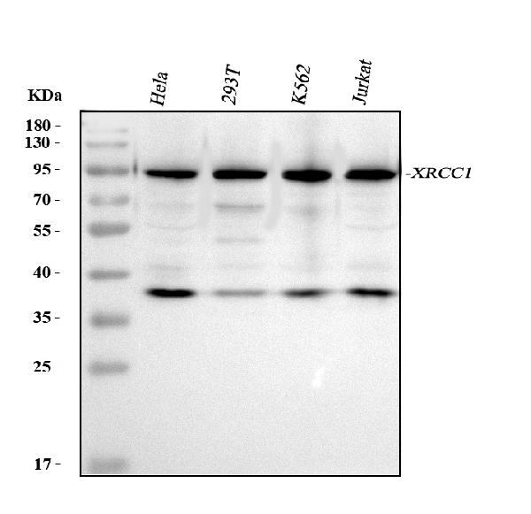
Facts about DNA repair protein XRCC1.
.
Human | |
---|---|
Gene Name: | XRCC1 |
Uniprot: | P18887 |
Entrez: | 7515 |
Belongs to: |
---|
No superfamily |
DNA repair protein XRCC1; RCC; X-ray repair complementing defective repair in Chinese hamster cells 1; X-ray repair cross-complementing protein 1; X-ray-repair, complementing defective, repair in Chinese hamster
Mass (kDA):
69.477 kDA
Human | |
---|---|
Location: | 19q13.31 |
Sequence: | 19; NC_000019.10 (43543311..43575527, complement) |
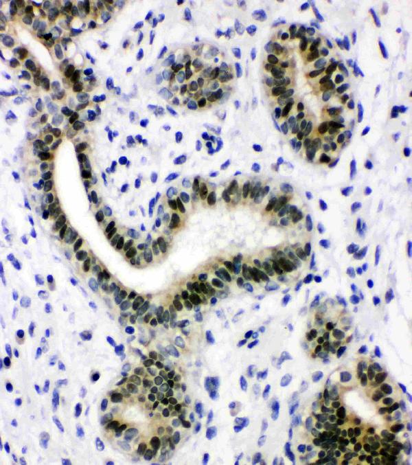
Expressed in fibroblasts, retinal pigmented epithelial cells and lymphoblastoid cells (at protein level).
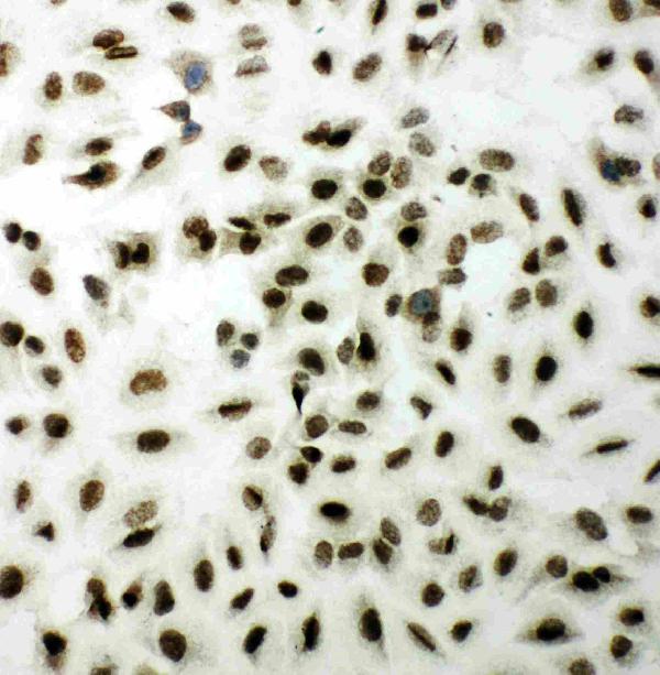
Nucleus. Moves from the nucleoli to the global nuclear chromatin upon DNA damage.
In this article, we will explore some of the best uses for the XRCC1 marker in a variety of contexts, including anti-Caveolin-1/CAV1, anti-p-Akt and anti-DNA repair protein XRCC3. The XRCC1 marker is also known as anti-Caveolin-1/Cav1.
XRCC1 is a genetic marker that can be used in molecular biology experiments. It can be used to measure the relative expression of XRCC1 in cells. It can be used to screen the effects of various treatments and activators. This kit is suitable for both human and animal samples. The antibody can be stored at -20°C for one year. It contains 5 mg BSA and 0.05 mg thimerosal.
The gene CAV1 provides instructions for the caveolin-1 protein, which appears to have multiple functions in various cells and tissues. Moreover, caveolin-1 promotes tumorigenesis in Apc(min/+) mice. Thus, the XRCC1 marker is a useful tool to monitor the level of caveolin-1 in human pancreatic cancer.
It has been shown that drug-resistant tumours have elevated levels of CAV1 expression. It has also been shown that stress can increase CAV1 expression in cancer cells. Hence, anti-neoplastic drugs administered at sub-cytotoxic levels may suffice to increase CAV1 expression in tumors. Further, the XRCC1 marker is an important tool for identifying the mechanisms underlying anti-Caveolin-1/CAV1 expression.
The role of CAV1 in cancer cell metabolism is still unclear. While the role of CAV1 in tumor metabolism is not completely understood, studies have revealed that CAV1 knockdown reduced glucose uptake, increased lactate output, and elevated levels of intracellular ATP. The CAV1 knockdown-induced autophagy also triggered AMPK-p53 signaling. Moreover, CAV1 interacts with insulin receptors and with the low-denser-related protein (LRP6). LRP6 stimulates IR kinases and enhances IR/IGF-1R signaling.
The Polymorphism of XRCC3 Thr241Met, which is associated with leukemia risk, has been a matter of intense study. The XRCC3 Thr241Met polymorphism is linked to an increased risk for leukemia in both the Caucasian and African-American population. Moreover, it has been associated with a higher risk of leukemia among whites and Spanish characters. To investigate the role of XRCC3 Thr241Met, two independent authors searched Pubmed, Embase, Cochrane library, Google academic, and China National Knowledge Infrastructure. The search time period is from the database's inception up to March 2021.
The Anti-DNA repair protein XRCC3, catalog # PA1697, is validated for use in WB and IHC assays. It reacts with Rat and Mouse. It contains 5 mg of BSA. In case of a high-quality sample, blocking peptide is also available for this antibody. The antibodies from Boster Bio have been approved for use in ELISA, flow cytometry, and IHC.
The XRCC3 protein has been shown to enhance the homologous recombination process in human brain cells. It interacts with HsRad51C, which is only found in tiny amounts in the brain. This protein binds human Rad51C, which exhibits the highest sequence identity. The XRCC3 protein is also linked to breast cancer and susceptibility to cutaneous malignant melanoma type 6.
The XRCC1 marker is a highly specific and sensitive method for the detection of mouse XRCC1. This antibody is a monoclonal antibody, suitable for detecting XRCC1 protein. XRCC proteins are responsible for efficiently repairing DNA base damage and maintaining genetic stability. These proteins share similar sequence with the yeast DNA repair protein Rad5. XRCC1 and XRCC2 are proteins involved in the base excision repair pathway and contribute to chromosome stability during cell division.
XRCC1 polymorphisms have several functions. They are involved in the regulation of gene expression and can either enhance or decrease the mutational response of RCC genes. Both XRCC1 and OGG1 polymorphisms affect methylation status. XRCC1 is involved in the methylation of p16 and TIMP3 and OGG1 increases the methylation of RCC gene expression.
The XRCC1 marker is associated with renal cell carcinoma (RCC). RCC is the most common malignant kidney tumor. It canonically activates the hypoxia-inducible transcription factor pathway. However, the mechanisms that underlie this pathway are poorly understood. Therefore, developing novel therapeutic strategies for RCC patients is of paramount importance. This study aims to evaluate the role of miR-183-5p in the development of RCC and how it might be used clinically in the future.
Researchers at the University of California, San Diego have developed a new detection system for snail mucin: the Anti-p-Snail Bostro Bio. This antibody uses a combination of two adjuvants to enhance neutrophil recruitment in snail mucus. This system also has a low background level and high specificity. Read on to learn more about this new detection system. Anti-p-Snail Boster Bio targets and binds to its target protein, reducing its toxicity and enhancing the detection of a wide range of protein types.
When snail mucin is combined with the rHBsAg vaccine, it produces significant increases in the number of monocytes. These cells are key players in activating adaptive immune responses and performing antimicrobial activities. Because these cells interact with antigen, snail mucin can enhance the immune response to the rHBsAg vaccine. Further molecular dynamics studies are necessary to assess this potential for vaccines.
In addition to its efficacy, SNHG1 knockdown has been found to suppress the expression of FOXK1 and MiR-376a in human tumor cells. This inhibits cell invasion and migration and promotes apoptosis. Snail knockdown is also associated with reduced expression of N-cadherin, Bax, and E-cadherin, which are important markers of cancer cells.
The XRCC1 gene is responsible for the production of a protein known as insulin. In vivo and in vitro, this protein phosphorylates CK2. It can be detected by aprataxin, a protein that binds to the XRCC1 gene through the FHA domain. The antibody is produced by Boster Bio.
The XRCC1 gene code for the human protein vimentin. Vimentin is expressed by human lymphocytes and Burkitt's lymphoma cells. It is also secreted by activated macrophages. The gene for human vimentin is located on the short arm of chromosome 10.
CircFLNA is an inhibitor of BCa cell proliferation, invasion, and migration. It also inhibits the expression of MMP-2 and vimentin. Furthermore, miR-216a-3p reverses the effect of circFLNA on protein expression. These results suggest that miR-216a-3p might regulate BCa malignancy by suppressing vimentin expression.
The antibodies were diluted 1:200 in DMSO and used as secondary antibodies. Secondary antibodies were rabbit anti-mouse IgG or horseradish-peroxidase-labeled goat anti-rabbit. Anti-cyclin E and p27 were used as controls. Anti-vimentin antibodies are available from the same suppliers.
PMID: 2247054 by Thompson L.H., et al. Molecular cloning of the human XRCC1 gene, which corrects defective DNA strand break repair and sister chromatid exchange.
PMID: 11163244 by Whitehouse C.J., et al. XRCC1 stimulates human polynucleotide kinase activity at damaged DNA termini and accelerates DNA single-strand break repair.
*More publications can be found for each product on its corresponding product page