Product Info Summary
| SKU: | PA1482 |
|---|---|
| Size: | 100 μg/vial |
| Reactive Species: | Human, Mouse, Rat |
| Host: | Rabbit |
| Application: | WB |
Customers Who Bought This Also Bought
Product info
Product Name
Anti-Tau/MAPT Antibody Picoband®
SKU/Catalog Number
PA1482
Size
100 μg/vial
Form
Lyophilized
Description
Boster Bio Anti-Tau/MAPT Antibody catalog # PA1482. Tested in WB applications. This antibody reacts with Human, Mouse, Rat. The brand Picoband indicates this is a premium antibody that guarantees superior quality, high affinity, and strong signals with minimal background in Western blot applications. Only our best-performing antibodies are designated as Picoband, ensuring unmatched performance.
Storage & Handling
Store at -20˚C for one year from date of receipt. After reconstitution, at 4˚C for one month. It can also be aliquotted and stored frozen at -20˚C for six months. Avoid repeated freeze-thaw cycles.
Cite This Product
Anti-Tau/MAPT Antibody Picoband® (Boster Biological Technology, Pleasanton CA, USA, Catalog # PA1482)
Host
Rabbit
Contents
Each vial contains 5mg BSA, 0.9mg NaCl, 0.2mg Na2HPO4, 0.05mg Thimerosal, 0.05mg NaN3.
Clonality
Polyclonal
Isotype
Rabbit IgG
Immunogen
A synthetic peptide corresponding to a sequence at the N-terminus of human Tau, different from the related mouse and rat sequences by three amino acids.
*Blocking peptide can be purchased. Costs vary based on immunogen length. Contact us for pricing.
Cross-reactivity
No cross-reactivity with other proteins
Reactive Species
PA1482 is reactive to MAPT in Human, Mouse, Rat
Reconstitution
Add 0.2ml of distilled water will yield a concentration of 500ug/ml.
Observed Molecular Weight
79 kDa
Calculated molecular weight
78928 MW
Background of Tau
MAPT, Microtubule-associated protein tau, appears to be enriched in axons. The MAPT gene is assigned to chromosome 17 by hybridization of a cDNA clone to flow-sorted and spot-blotted chromosomes and to 17q21 by in situ hybridization, containing 16 exons. The tau proteins are the product of alternative splicingfrom a single gene that in humans is designated MAPT. Tau proteins are proteins that stabilize microtubules. They are abundant in neurons in the central nervous system and are less common elsewhere. When tau proteins are defective, and no longer stabilize microtubules properly, they can result in dementias such as Alzheimer's disease.
Antibody Validation
Boster validates all antibodies on WB, IHC, ICC, Immunofluorescence, and ELISA with known positive control and negative samples to ensure specificity and high affinity, including thorough antibody incubations.
Application & Images
Applications
PA1482 is guaranteed for WB Boster Guarantee
Assay Dilutions Recommendation
The recommendations below provide a starting point for assay optimization. The actual working concentration varies and should be decided by the user.
Western blot, 0.1-0.5μg/ml, Human, Mouse, Rat
Positive Control
WB: Rat Brain Tissue, Mouse Brain Tissue, HT1080 Whole Cell, MCF-7 Whole Cell
Validation Images & Assay Conditions

Click image to see more details
Anti-Tau antibody, PA1482, Western blotting
All lanes: Anti (PA1482) at 0.5ug/ml
Lane 1: Rat Brain Tissue Lysate at 50ug
Lane 2: Mouse Brain Tissue Lysate at 50ug
Lane 3: HT1080 Whole Cell Lysate at 40ug
Lane 4: MCF-7 Whole Cell Lysate at 40ug
Predicted bind size: 79KD
Observed bind size: 79KD
Protein Target Info & Infographic
Gene/Protein Information For MAPT (Source: Uniprot.org, NCBI)
Gene Name
MAPT
Full Name
Microtubule-associated protein tau
Weight
78928 MW
Alternative Names
Microtubule-associated protein tau;Neurofibrillary tangle protein;Paired helical filament-tau;PHF-tau;MAPT;MAPTL, MTBT1, TAU; MAPT DDPAC, FTDP-17L, MSTD, MTBT1, MTBT2, PPND, PPP1R103, TAU, tau-40, MAPT microtubule associated protein tau microtubule-associated protein tau|G protein beta1/gamma2 subunit-interacting factor 1|PHF-tau|neurofibrillary tangle protein|paired helical filament-tau|protein phosphatase 1, regulatory subunit 103
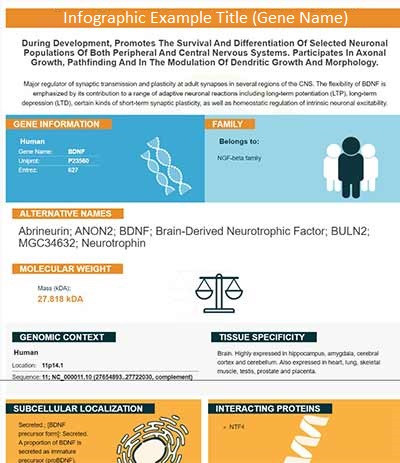
*If product is indicated to react with multiple species, protein info is based on the gene entry specified above in "Species".For more info on MAPT, check out the MAPT Infographic

We have 30,000+ of these available, one for each gene! Check them out.
In this infographic, you will see the following information for MAPT: database IDs, superfamily, protein function, synonyms, molecular weight, chromosomal locations, tissues of expression, subcellular locations, post-translational modifications, and related diseases, research areas & pathways. If you want to see more information included, or would like to contribute to it and be acknowledged, please contact [email protected].
Specific Publications For Anti-Tau/MAPT Antibody Picoband® (PA1482)
Hello CJ!
PA1482 has been cited in 10 publications:
*The publications in this section are manually curated by our staff scientists. They may differ from Bioz's machine gathered results. Both are accurate. If you find a publication citing this product but is missing from this list, please let us know we will issue you a thank-you coupon.
Micro-RNA-137 Inhibits Tau Hyperphosphorylation in Alzheimer's Disease and Targets the CACNA1C Gene in Transgenic Mice and Human Neuroblastoma %u2026
Lychee seed extract protects against neuronal injury and improves cognitive function in rats with type II diabetes mellitus with cognitive impairment
The expression of insulin-like growth factor-1 in senior patients with diabetes and dementia
Panax notoginsenoside Rb1 ameliorates Alzheimer?s disease by upregulating brain-derived neurotrophic factor and downregulating Tau protein expression
Chen C, Gu J, Basurto-Islas G, Jin N, Wu F, Gong CX, Iqbal K, Liu F. Sci Rep. 2017 Oct 18;7(1):13478. doi: 10.1038/s41598-017-13791-5. Up-regulation of casein kinase 1ε is involved in tau pathogenesis in Alzheimer’s disease
Han F1, Zhuang TT1, Chen JJ1, Zhu XL1,2, Cai YF1, Lu YP1. PLoS One. 2017 Sep 21;12(9):e0185102. doi: 10.1371/journal.pone.0185102. eCollection 2017. Novel derivative of Paeonol, Paeononlsilatie sodium, alleviates behavioral damage and hippocampal ...
Li X, Li M, Li Y, Quan Q, Wang J. Neural Regen Res. 2012 Dec 25;7(36):2860-6. Doi: 10.3969/J.Issn.1673-5374.2012.36.002. Cellular And Molecular Mechanisms Underlying The Action Of Ginsenoside Rg1 Against Alzheimer'S Disease.
Zhu Yf, Li Xh, Yuan Zp, Li Cy, Tian Rb, Jia W, Xiao Zp. Eur J Pharmacol. 2015 Sep 5;762:239-46. Doi: 10.1016/J.Ejphar.2015.06.002. Epub 2015 Jun 3. Allicin Improves Endoplasmic Reticulum Stress-Related Cognitive Deficits Via Perk/Nrf2 Antioxidativ...
Tan Wf, Cao Xz, Wang Jk, Lv Hw, Wu By, Ma H. Clin Exp Pharmacol Physiol. 2010 Oct;37(10):1010-5. Doi: 10.1111/J.1440-1681.2010.05433.X. Protective Effects Of Lithium Treatment For Spatial Memory Deficits Induced By Tau Hyperphosphorylation In Sple...
Tu Q, Pang L, Chen Y, Zhang Y, Zhang R, Lu B, Wang J. Analyst. 2014 Jan 7;139(1):105-15. Doi: 10.1039/C3An01796F. Epub 2013 Oct 25. Effects Of Surface Charges Of Graphene Oxide On Neuronal Outgrowth And Branching.
Recommended Resources
Here are featured tools and databases that you might find useful.
- Boster's Pathways Library
- Protein Databases
- Bioscience Research Protocol Resources
- Data Processing & Analysis Software
- Photo Editing Software
- Scientific Literature Resources
- Research Paper Management Tools
- Molecular Biology Software
- Primer Design Tools
- Bioinformatics Tools
- Phylogenetic Tree Analysis
Customer Reviews
Have you used Anti-Tau/MAPT Antibody Picoband®?
Submit a review and receive an Amazon gift card.
- $30 for a review with an image
0 Reviews For Anti-Tau/MAPT Antibody Picoband®
Customer Q&As
Have a question?
Find answers in Q&As, reviews.
Can't find your answer?
Submit your question
4 Customer Q&As for Anti-Tau/MAPT Antibody Picoband®
Question
We are currently using anti-Tau/MAPT antibody PA1482 for mouse tissue, and we are happy with the WB results. The species of reactivity given in the datasheet says human, mouse, rat. Is it possible that the antibody can work on primate tissues as well?
Verified Customer
Verified customer
Asked: 2019-01-02
Answer
The anti-Tau/MAPT antibody (PA1482) has not been tested for cross reactivity specifically with primate tissues, though there is a good chance of cross reactivity. We have an innovator award program that if you test this antibody and show it works in primate you can get your next antibody for free. Please contact me if I can help you with anything.
Boster Scientific Support
Answered: 2019-01-02
Question
We were well pleased with the WB result of your anti-Tau/MAPT antibody. However we have been able to see positive staining in cervix carcinoma erythroleukemia cytosol using this antibody. Is that expected? Could you tell me where is MAPT supposed to be expressed?
Verified Customer
Verified customer
Asked: 2018-12-10
Answer
From literature, cervix carcinoma erythroleukemia does express MAPT. Generally MAPT expresses in cytoplasm, cytosol. Regarding which tissues have MAPT expression, here are a few articles citing expression in various tissues:
Brain, Pubmed ID: 1512244, 2484340, 2495000, 2498079, 3131773, 15489334
Cervix carcinoma, Pubmed ID: 16964243, 18220336, 20068231
Cervix carcinoma, and Erythroleukemia, Pubmed ID: 23186163
Fetal brain, Pubmed ID: 2516729
Fetal brain cortex, Pubmed ID: 16443603
Leukemic T-cell, Pubmed ID: 19690332
Liver, Pubmed ID: 24275569
Boster Scientific Support
Answered: 2018-12-10
Question
you antibody using your anti-Tau/MAPT antibody for axonal transport of mitochondrion studies. Has this antibody been tested with western blotting on rat brain tissue? We would like to see some validation images before ordering.
Verified Customer
Verified customer
Asked: 2018-07-12
Answer
We appreciate your inquiry. This PA1482 anti-Tau/MAPT antibody is tested on rat brain tissue, tissue lysate, mouse brain, ht1080 whole cell lysate. It is guaranteed to work for WB in human, mouse, rat. Our Boster guarantee will cover your intended experiment even if the sample type has not been be directly tested.
Boster Scientific Support
Answered: 2018-07-12
Question
We have observed staining in mouse brain. Are there any suggestions? Is anti-Tau/MAPT antibody supposed to stain brain positively?
L. Mitchell
Verified customer
Asked: 2014-10-06
Answer
From what I have seen in literature brain does express MAPT. From what I have seen in Uniprot.org, MAPT is expressed in parietal lobe, brain, fetal brain, fetal brain cortex, cervix carcinoma, leukemic t-cell, cervix carcinoma erythroleukemia, liver, among other tissues. Regarding which tissues have MAPT expression, here are a few articles citing expression in various tissues:
Brain, Pubmed ID: 1512244, 2484340, 2495000, 2498079, 3131773, 15489334
Cervix carcinoma, Pubmed ID: 16964243, 18220336, 20068231
Cervix carcinoma, and Erythroleukemia, Pubmed ID: 23186163
Fetal brain, Pubmed ID: 2516729
Fetal brain cortex, Pubmed ID: 16443603
Leukemic T-cell, Pubmed ID: 19690332
Liver, Pubmed ID: 24275569
Boster Scientific Support
Answered: 2014-10-06


