Click image to see more details
Product Info Summary
| SKU: | A07697-1 |
|---|---|
| Size: | 100 µg/vial |
| Reactive Species: | Human |
| Host: | Rabbit |
| Application: | ELISA, IF, ICC, WB |
Customers Who Bought This Also Bought


Product info
Product Name
Anti-PSMB2 Antibody Picoband®
View all Proteasome 20S beta2 Antibodies
SKU/Catalog Number
A07697-1
Size
100 µg/vial
Form
Lyophilized
Description
Boster Bio Anti-PSMB2 Antibody Picoband® catalog # A07697-1. Tested in ELISA, IF, ICC, WB applications. This antibody reacts with Human. The brand Picoband indicates this is a premium antibody that guarantees superior quality, high affinity, and strong signals with minimal background in Western blot applications. Only our best-performing antibodies are designated as Picoband, ensuring unmatched performance.
Storage & Handling
At -20°C for one year from date of receipt. After reconstitution, at 4°C for one month. It can also be aliquotted and stored frozen at -20°C for six months. Avoid repeated freezing and thawing.
Cite This Product
Anti-PSMB2 Antibody Picoband® (Boster Biological Technology, Pleasanton CA, USA, Catalog # A07697-1)
Host
Rabbit
Contents
Each vial contains 4 mg Trehalose, 0.9 mg NaCl, 0.2 mg Na2HPO4.
Clonality
Polyclonal
Isotype
Rabbit IgG
Immunogen
E.coli-derived human PSMB2 recombinant protein (Position: M1-S201).
*Blocking peptide can be purchased. Costs vary based on immunogen length. Contact us for pricing.
Cross-reactivity
No cross-reactivity with other proteins.
Reactive Species
A07697-1 is reactive to PSMB2 in Human
Reconstitution
Adding 0.2 ml of distilled water will yield a concentration of 500 µg/ml.
Observed Molecular Weight
23 kDa
Calculated molecular weight
23 kDa
Background of Proteasome 20S beta2
Proteasome subunit beta type-2 also known as 20S proteasome subunit beta-4 (based on systematic nomenclature) is a protein that in humans is encoded by the PSMB2 gene. The proteasome is a multicatalytic proteinase complex with a highly ordered ring-shaped 20S core structure. The core structure is composed of 4 rings of 28 non-identical subunits; 2 rings are composed of 7 alpha subunits and 2 rings are composed of 7 beta subunits. Proteasomes are distributed throughout eukaryotic cells at a high concentration and cleave peptides in an ATP/ubiquitin-dependent process in a non-lysosomal pathway. An essential function of a modified proteasome, the immunoproteasome, is the processing of class I MHC peptides. This gene encodes a member of the proteasome B-type family, also known as the T1B family, that is a 20S core beta subunit. Multiple alternatively spliced transcript variants encoding distinct isoforms have been found for this gene.
Antibody Validation
Boster validates all antibodies on WB, IHC, ICC, Immunofluorescence, and ELISA with known positive control and negative samples to ensure specificity and high affinity, including thorough antibody incubations.
Application & Images
Applications
A07697-1 is guaranteed for ELISA, IF, ICC, WB Boster Guarantee
Assay Dilutions Recommendation
The recommendations below provide a starting point for assay optimization. The actual working concentration varies and should be decided by the user.
Western blot, 0.25-0.5 μg/ml, Human
Immunocytochemistry/Immunofluorescence, 5 μg/ml, Human
ELISA, 0.1-0.5 μg/ml, -
Positive Control
WB: human 293T whole cell, human Hela whole cell
ICC/IF: Hela cell
Validation Images & Assay Conditions
Click image to see more details
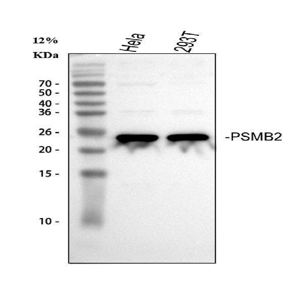
Figure 1. Western blot analysis of PSMB2 using anti-PSMB2 antibody (A07697-1).
Electrophoresis was performed on a 5-20% SDS-PAGE gel at 70V (Stacking gel) / 90V (Resolving gel) for 2-3 hours. The sample well of each lane was loaded with 30 ug of sample under reducing conditions.
Lane 1: human Hela whole cell lysates,
Lane 2: human 293T whole cell lysates.
After electrophoresis, proteins were transferred to a nitrocellulose membrane at 150 mA for 50-90 minutes. Blocked the membrane with 5% non-fat milk/TBS for 1.5 hour at RT. The membrane was incubated with rabbit anti-PSMB2 antigen affinity purified polyclonal antibody (Catalog # A07697-1) at 0.5 μg/mL overnight at 4°C, then washed with TBS-0.1%Tween 3 times with 5 minutes each and probed with a goat anti-rabbit IgG-HRP secondary antibody at a dilution of 1:5000 for 1.5 hour at RT. The signal is developed using an Enhanced Chemiluminescent detection (ECL) kit (Catalog # EK1002) with Tanon 5200 system. A specific band was detected for PSMB2 at approximately 23 kDa. The expected band size for PSMB2 is at 23 kDa.

Click image to see more details
Figure 2. IF analysis of PSMB2 using anti-PSMB2 antibody (A07697-1).
PSMB2 was detected in an immunocytochemical section of Hela cells. Enzyme antigen retrieval was performed using IHC enzyme antigen retrieval reagent (AR0022) for 15 mins. The cells were blocked with 10% goat serum. And then incubated with 5 μg/mL rabbit anti-PSMB2 Antibody (A07697-1) overnight at 4°C. Cy3 Conjugated Goat Anti-Rabbit IgG (BA1032) was used as secondary antibody at 1:500 dilution and incubated for 30 minutes at 37°C. The section was counterstained with DAPI. Visualize using a fluorescence microscope and filter sets appropriate for the label used.
Protein Target Info & Infographic
Gene/Protein Information For PSMB2 (Source: Uniprot.org, NCBI)
Gene Name
PSMB2
Full Name
Proteasome subunit beta type-2
Weight
23 kDa
Superfamily
peptidase T1B family
Alternative Names
Chromaffin granule amine transporter; Solute carrier family 18 member 1; Vesicular amine transporter 1; VAT1; SLC18A1; VAT1; VMAT1 PSMB2 HC7-I proteasome 20S subunit beta 2 proteasome subunit beta type-2|macropain subunit C7-I|multicatalytic endopeptidase complex subunit C7-1|multicatalytic endopeptidase complex subunit C7-I|proteasome (prosome, macropain) subunit, beta type, 2|proteasome beta 2 subunit|proteasome component C7-I|proteasome subunit beta 2|proteasome subunit beta4|proteasome subunit, beta type, 2|testicular tissue protein Li 152
*If product is indicated to react with multiple species, protein info is based on the gene entry specified above in "Species".For more info on PSMB2, check out the PSMB2 Infographic

We have 30,000+ of these available, one for each gene! Check them out.
In this infographic, you will see the following information for PSMB2: database IDs, superfamily, protein function, synonyms, molecular weight, chromosomal locations, tissues of expression, subcellular locations, post-translational modifications, and related diseases, research areas & pathways. If you want to see more information included, or would like to contribute to it and be acknowledged, please contact [email protected].
Specific Publications For Anti-PSMB2 Antibody Picoband® (A07697-1)
Loading publications
Recommended Resources
Here are featured tools and databases that you might find useful.
- Boster's Pathways Library
- Protein Databases
- Bioscience Research Protocol Resources
- Data Processing & Analysis Software
- Photo Editing Software
- Scientific Literature Resources
- Research Paper Management Tools
- Molecular Biology Software
- Primer Design Tools
- Bioinformatics Tools
- Phylogenetic Tree Analysis
Customer Reviews
Have you used Anti-PSMB2 Antibody Picoband®?
Submit a review and receive an Amazon gift card.
- $30 for a review with an image
0 Reviews For Anti-PSMB2 Antibody Picoband®
Customer Q&As
Have a question?
Find answers in Q&As, reviews.
Can't find your answer?
Submit your question
