Product Info Summary
| SKU: | M00413 |
|---|---|
| Size: | 100ug/vial |
| Reactive Species: | Cow, Dog, Goat, Guinea pig, Human, Monkey, Mouse, Pig, Rabbit, Rat, Horse, Cat, Baboon |
| Host: | Mouse |
| Application: | Flow Cytometry, IF, IHC |
Customers Who Bought This Also Bought
Product info
Product Name
Anti-Macrophage L1 Protein S100A8 Monoclonal Antibody
View all S100A8 & S100A9 Antibodies
SKU/Catalog Number
M00413
Size
100ug/vial
Form
Liquid
Description
Boster Bio Anti-Macrophage L1 Protein S100A8 Monoclonal Antibody (Catalog # M00413). Tested in Flow Cytometry, IF, IHC applications. This antibody reacts with Human, Baboon, Monkey, Cow, Pig, Goat, Horse, Cat, Dog, Rabbit, Guinea pig, Rat, Mouse.
Storage & Handling
Antibody with azide - store at 2 to 8°C. Antibody without azide - store at -20 to -80°C. Antibody is stable for 24 months. Non-hazardous. No MSDS required.
Cite This Product
Anti-Macrophage L1 Protein S100A8 Monoclonal Antibody (Boster Biological Technology, Pleasanton CA, USA, Catalog # M00413)
Host
Mouse
Contents
Prepared in 10mM PBS with 0.05% BSA & 0.05% azide. Also available WITHOUT BSA & azide at 1.0mg/ml.
Clonality
Monoclonal
Clone Number
Clone: SPM281
Isotype
IgG1, kappa
Immunogen
Affinity Purified monocyte membrane preparation
*Blocking peptide can be purchased. Costs vary based on immunogen length. Contact us for pricing.
Reactive Species
M00413 is reactive to S100A8 & S100A9 in Cow, Dog, Goat, Guinea pig, Human, Monkey, Mouse, Pig, Rabbit, Rat, Horse, Cat, Baboon
Reconstitution
Reconstitute with 1ml water (15 minutes at room temperature).
Calculated molecular weight
10835 MW
Background of S100A8 & S100A9
Recognizes the L1 or Calprotectin molecule, an intra-cytoplasmic antigen comprising of a 12kDa alpha chain and a 14kDa beta chain expressed by granulocytes, monocytes and by tissue macrophages. Macrophages usually arise from hematopoietic stem cells in the bone marrow. Under migration into tissues, the monocytes undergo further differentiation to become multifunctional tissue macrophages. They are classified into normal and inflammatory macrophages. Normal macrophages include macrophages in connective tissue (histiocytes), liver (Kupffer's cells), lung (alveolar macrophages), lymph nodes (free and fixed macrophages), spleen (free and fixed macrophages), bone marrow (fixed macrophages), serous fluids (pleural and peritoneal macrophages), skin (histiocytes, Langerhans's cell) and in other tissues. Inflammatory macrophages are present in various exudates. Macrophages are part of the innate immune system, recognizing, engulfing and destroying many potential pathogens including bacteria, pathogenic protozoa, fungi and helminthes. This monoclonal antibody reacts with neutrophils, monocytes, macrophages, and squamous mucosal epithelia and has been shown as an important marker for identifying macrophages in tissue sections.
Antibody Validation
Boster validates all antibodies on WB, IHC, ICC, Immunofluorescence, and ELISA with known positive control and negative samples to ensure specificity and high affinity, including thorough antibody incubations.
Application & Images
Applications
M00413 is guaranteed for Flow Cytometry, IF, IHC Boster Guarantee
Assay Dilutions Recommendation
The recommendations below provide a starting point for assay optimization. The actual working concentration varies and should be decided by the user.
Flow Cytometry (1-2ug/million cells)
Immunofluorescence (1-2ug/ml)
Immunohistochemistry (Formalin-fixed) (1-2ug/ml for 30 minutes at RT)(Staining of formalin-fixed tissues requires heating tissue sections in 10mM Tris with 1mM EDTA, pH 9.0, for 45 min at 95°C followed by cooling at RT for 20 minutes)
Optimal dilution for a specific application should be determined.
Validation Images & Assay Conditions

Click image to see more details
Formalin-fixed, paraffin-embedded human Tonsil stained with Anti-Macrophage L1 Protein Monoclonal Antibody (SPM281)
Protein Target Info & Infographic
Gene/Protein Information For S100A8 & S100A9 (Source: Uniprot.org, NCBI)
Gene Name
S100A8 & S100A9
Full Name
Weight
10835 MW
Alternative Names
Protein S100-A8;Calgranulin-A;Calprotectin L1L subunit;Cystic fibrosis antigen;CFAG;Leukocyte L1 complex light chain;Migration inhibitory factor-related protein 8;MRP-8;p8;S100 calcium-binding protein A8;Urinary stone protein band A;Protein S100-A8, N-terminally processed;S100A8;CAGA, CFAG, MRP8;
*If product is indicated to react with multiple species, protein info is based on the gene entry specified above in "Species".For more info on S100A8 & S100A9, check out the S100A8 & S100A9 Infographic

We have 30,000+ of these available, one for each gene! Check them out.
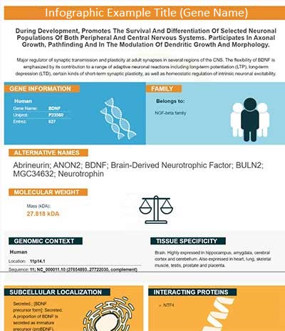
In this infographic, you will see the following information for S100A8 & S100A9: database IDs, superfamily, protein function, synonyms, molecular weight, chromosomal locations, tissues of expression, subcellular locations, post-translational modifications, and related diseases, research areas & pathways. If you want to see more information included, or would like to contribute to it and be acknowledged, please contact [email protected].
Specific Publications For Anti-Macrophage L1 Protein S100A8 Monoclonal Antibody (M00413)
Hello CJ!
No publications found for M00413
*Do you have publications using this product? Share with us and receive a reward. Ask us for more details.
Recommended Resources
Here are featured tools and databases that you might find useful.
- Boster's Pathways Library
- Protein Databases
- Bioscience Research Protocol Resources
- Data Processing & Analysis Software
- Photo Editing Software
- Scientific Literature Resources
- Research Paper Management Tools
- Molecular Biology Software
- Primer Design Tools
- Bioinformatics Tools
- Phylogenetic Tree Analysis
Customer Reviews
Have you used Anti-Macrophage L1 Protein S100A8 Monoclonal Antibody?
Submit a review and receive an Amazon gift card.
- $30 for a review with an image
0 Reviews For Anti-Macrophage L1 Protein S100A8 Monoclonal Antibody
Customer Q&As
Have a question?
Find answers in Q&As, reviews.
Can't find your answer?
Submit your question
3 Customer Q&As for Anti-Macrophage L1 Protein S100A8 Monoclonal Antibody
Question
We were content with the WB result of your anti-Macrophage L1 Protein Monoclonal antibody. However we have seen positive staining in neutrophil secreted. cytoplasm. cytoplasm, using this antibody. Is that expected? Could you tell me where is S100A8 supposed to be expressed?
Verified Customer
Verified customer
Asked: 2019-07-30
Answer
According to literature, neutrophil does express S100A8. Generally S100A8 expresses in secreted. cytoplasm. cytoplasm,. Regarding which tissues have S100A8 expression, here are a few articles citing expression in various tissues:
Ascites, Pubmed ID: 7695842
Keratinocyte, Pubmed ID: 1286667
Liver, Pubmed ID: 24275569
Neutrophil, Pubmed ID: 1326551
Skeletal muscle, Pubmed ID: 15489334
Tongue, Pubmed ID: 14702039
Boster Scientific Support
Answered: 2019-07-30
Question
We have seen staining in monkey keratinocyte. What should we do? Is anti-Macrophage L1 Protein Monoclonal antibody supposed to stain keratinocyte positively?
Verified Customer
Verified customer
Asked: 2019-06-07
Answer
According to literature keratinocyte does express S100A8. According to Uniprot.org, S100A8 is expressed in pharyngeal mucosa, tongue, skeletal muscle, neutrophil, ascites, keratinocyte, liver, among other tissues. Regarding which tissues have S100A8 expression, here are a few articles citing expression in various tissues:
Ascites, Pubmed ID: 7695842
Keratinocyte, Pubmed ID: 1286667
Liver, Pubmed ID: 24275569
Neutrophil, Pubmed ID: 1326551
Skeletal muscle, Pubmed ID: 15489334
Tongue, Pubmed ID: 14702039
Boster Scientific Support
Answered: 2019-06-07
Question
We are currently using anti-Macrophage L1 Protein Monoclonal antibody M00413 for goat tissue, and we are well pleased with the IHC results. The species of reactivity given in the datasheet says bovine, canine, equine, goat, guinea pig, human, monkey, pig, rabbit, rat. Is it possible that the antibody can work on feline tissues as well?
K. Singh
Verified customer
Asked: 2014-04-08
Answer
The anti-Macrophage L1 Protein Monoclonal antibody (M00413) has not been tested for cross reactivity specifically with feline tissues, though there is a good chance of cross reactivity. We have an innovator award program that if you test this antibody and show it works in feline you can get your next antibody for free. Please contact me if I can help you with anything.
Boster Scientific Support
Answered: 2014-04-08


