Click image to see more details
-

-
-
-
-
+6
Product Info Summary
| SKU: | A11090-1 |
|---|---|
| Size: | 100 μg/vial |
| Reactive Species: | Human |
| Host: | Rabbit |
| Application: | ELISA, Flow Cytometry, IF, IHC, ICC, WB |
Customers Who Bought This Also Bought

Product info
Product Name
Anti-HNRNPH3 Antibody Picoband®
SKU/Catalog Number
A11090-1
Size
100 μg/vial
Form
Lyophilized
Description
Boster Bio Anti-HNRNPH3 Antibody Picoband® catalog # A11090-1. Tested in ELISA, Flow Cytometry, IF, IHC, ICC, WB applications. This antibody reacts with Human. The brand Picoband indicates this is a premium antibody that guarantees superior quality, high affinity, and strong signals with minimal background in Western blot applications. Only our best-performing antibodies are designated as Picoband, ensuring unmatched performance.
Storage & Handling
Store at -20˚C for one year from date of receipt. After reconstitution, at 4˚C for one month. It can also be aliquotted and stored frozen at -20˚C for six months. Avoid repeated freeze-thaw cycles.
Cite This Product
Anti-HNRNPH3 Antibody Picoband® (Boster Biological Technology, Pleasanton CA, USA, Catalog # A11090-1)
Host
Rabbit
Contents
Each vial contains 4mg Trehalose, 0.9mg NaCl and 0.2mg Na2HPO4.
Clonality
Polyclonal
Isotype
Rabbit IgG
Immunogen
E.coli-derived human HNRNPH3 recombinant protein (Position: K29-N268).
*Blocking peptide can be purchased. Costs vary based on immunogen length. Contact us for pricing.
Cross-reactivity
No cross-reactivity with other proteins.
Reactive Species
A11090-1 is reactive to HNRNPH3 in Human
Reconstitution
Add 0.2ml of distilled water will yield a concentration of 500ug/ml.
Observed Molecular Weight
35 kDa, 37 kDa
Calculated molecular weight
36.926kDa
Background of HNRPH3
Heterogeneous nuclear ribonucleoprotein H3 is a protein that in humans is encoded by the HNRNPH3 gene. This gene belongs to the subfamily of ubiquitously expressed heterogeneous nuclear ribonucleoproteins (hnRNPs). The hnRNPs are RNA binding proteins and they complex with heterogeneous nuclear RNA (hnRNA). These proteins are associated with pre-mRNAs in the nucleus and appear to influence pre-mRNA processing and other aspects of mRNA metabolism and transport. While all of the hnRNPs are present in the nucleus, some seem to shuttle between the nucleus and the cytoplasm. The hnRNP proteins have distinct nucleic acid binding properties. The protein encoded by this gene has two repeats of quasi-RRM domains that bind to RNAs. It is localized in nuclear bodies of the nucleus. This protein is involved in the splicing process and it also participates in early heat shock-induced splicing arrest by transiently leaving the hnRNP complexes. Several alternatively spliced transcript variants have been noted for this gene, however, not all are fully characterized.
Antibody Validation
Boster validates all antibodies on WB, IHC, ICC, Immunofluorescence, and ELISA with known positive control and negative samples to ensure specificity and high affinity, including thorough antibody incubations.
Application & Images
Applications
A11090-1 is guaranteed for ELISA, Flow Cytometry, IF, IHC, ICC, WB Boster Guarantee
Assay Dilutions Recommendation
The recommendations below provide a starting point for assay optimization. The actual working concentration varies and should be decided by the user.
Western blot, 0.25-0.5μg/ml, Human
Immunohistochemistry (Paraffin-embedded Section), 1-2μg/ml, Human
Immunocytochemistry/Immunofluorescence, 5μg/ml, Human
Flow Cytometry (Fixed), 1-3μg/1x106 cells, Human
ELISA, 0.1-0.5μg/ml, Human,
Positive Control
WB: human Jurkat whole cell, human PC-3 whole cell, human Raji whole cell, human K562 whole cell, human Caco-2 whole cell, human MCF-7 whole cell, human HL-60 whole cell, human PC-3 whole cell
IHC: human breast cancer tissue, human gastric cancer lymph node tissue, human liver cancer tissue, human ovarian serous adenocarcinoma tissue, human pancreatic cancer tissue, human placenta tissue, human rectal cancer tissue
ICC/IF: MCF-7 cell
FCM: K562 cell
Validation Images & Assay Conditions
Click image to see more details
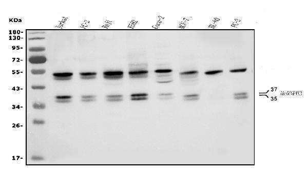
Figure 1. Western blot analysis of HNRNPH3 using anti-HNRNPH3 antibody (A11090-1).
Electrophoresis was performed on a 5-20% SDS-PAGE gel at 70V (Stacking gel) / 90V (Resolving gel) for 2-3 hours. The sample well of each lane was loaded with 30ug of sample under reducing conditions.
Lane 1: human Jurkat whole cell lysates,
Lane 2: human PC-3 whole cell lysates,
Lane 3: human Raji whole cell lysates,
Lane 4: human K562 whole cell lysates,
Lane 5: human Caco-2 whole cell lysates,
Lane 6: human MCF-7 whole cell lysates,
Lane 7: human HL-60 whole cell lysates,
Lane 8: human PC-3 whole cell lysates.
After Electrophoresis, proteins were transferred to a Nitrocellulose membrane at 150mA for 50-90 minutes. Blocked the membrane with 5% Non-fat Milk/ TBS for 1.5 hour at RT. The membrane was incubated with rabbit anti-HNRNPH3 antigen affinity purified polyclonal antibody (Catalog # A11090-1) at 0.5 μg/mL overnight at 4°C, then washed with TBS-0.1%Tween 3 times with 5 minutes each and probed with a goat anti-rabbit IgG-HRP secondary antibody at a dilution of 1:5000 for 1.5 hour at RT. The signal is developed using an Enhanced Chemiluminescent detection (ECL) kit (Catalog # EK1002) with Tanon 5200 system. A specific band was detected for HNRNPH3 at approximately 35KD and 37KD. The expected band size for HNRNPH3 is at 35KD and 37KD.

Click image to see more details
Figure 2. IHC analysis of HNRNPH3 using anti-HNRNPH3 antibody (A11090-1).
HNRNPH3 was detected in paraffin-embedded section of human breast cancer tissue. Heat mediated antigen retrieval was performed in EDTA buffer (pH8.0, epitope retrieval solution). The tissue section was blocked with 10% goat serum. The tissue section was then incubated with 2μg/ml rabbit anti-HNRNPH3 Antibody (A11090-1) overnight at 4°C. Biotinylated goat anti-rabbit IgG was used as secondary antibody and incubated for 30 minutes at 37°C. The tissue section was developed using Strepavidin-Biotin-Complex (SABC) (Catalog # SA1022) with DAB as the chromogen.

Click image to see more details
Figure 3. IHC analysis of HNRNPH3 using anti-HNRNPH3 antibody (A11090-1).
HNRNPH3 was detected in paraffin-embedded section of human gastric cancer lymph node tissue. Heat mediated antigen retrieval was performed in EDTA buffer (pH8.0, epitope retrieval solution). The tissue section was blocked with 10% goat serum. The tissue section was then incubated with 2μg/ml rabbit anti-HNRNPH3 Antibody (A11090-1) overnight at 4°C. Biotinylated goat anti-rabbit IgG was used as secondary antibody and incubated for 30 minutes at 37°C. The tissue section was developed using Strepavidin-Biotin-Complex (SABC) (Catalog # SA1022) with DAB as the chromogen.

Click image to see more details
Figure 4. IHC analysis of HNRNPH3 using anti-HNRNPH3 antibody (A11090-1).
HNRNPH3 was detected in paraffin-embedded section of human liver cancer tissue. Heat mediated antigen retrieval was performed in EDTA buffer (pH8.0, epitope retrieval solution). The tissue section was blocked with 10% goat serum. The tissue section was then incubated with 2μg/ml rabbit anti-HNRNPH3 Antibody (A11090-1) overnight at 4°C. Biotinylated goat anti-rabbit IgG was used as secondary antibody and incubated for 30 minutes at 37°C. The tissue section was developed using Strepavidin-Biotin-Complex (SABC) (Catalog # SA1022) with DAB as the chromogen.

Click image to see more details
Figure 5. IHC analysis of HNRNPH3 using anti-HNRNPH3 antibody (A11090-1).
HNRNPH3 was detected in paraffin-embedded section of human ovarian serous adenocarcinoma tissue. Heat mediated antigen retrieval was performed in EDTA buffer (pH8.0, epitope retrieval solution). The tissue section was blocked with 10% goat serum. The tissue section was then incubated with 2μg/ml rabbit anti-HNRNPH3 Antibody (A11090-1) overnight at 4°C. Biotinylated goat anti-rabbit IgG was used as secondary antibody and incubated for 30 minutes at 37°C. The tissue section was developed using Strepavidin-Biotin-Complex (SABC) (Catalog # SA1022) with DAB as the chromogen.

Click image to see more details
Figure 6. IHC analysis of HNRNPH3 using anti-HNRNPH3 antibody (A11090-1).
HNRNPH3 was detected in paraffin-embedded section of human pancreatic cancer tissue. Heat mediated antigen retrieval was performed in EDTA buffer (pH8.0, epitope retrieval solution). The tissue section was blocked with 10% goat serum. The tissue section was then incubated with 2μg/ml rabbit anti-HNRNPH3 Antibody (A11090-1) overnight at 4°C. Biotinylated goat anti-rabbit IgG was used as secondary antibody and incubated for 30 minutes at 37°C. The tissue section was developed using Strepavidin-Biotin-Complex (SABC) (Catalog # SA1022) with DAB as the chromogen.

Click image to see more details
Figure 7. IHC analysis of HNRNPH3 using anti-HNRNPH3 antibody (A11090-1).
HNRNPH3 was detected in paraffin-embedded section of human placenta tissue. Heat mediated antigen retrieval was performed in EDTA buffer (pH8.0, epitope retrieval solution). The tissue section was blocked with 10% goat serum. The tissue section was then incubated with 2μg/ml rabbit anti-HNRNPH3 Antibody (A11090-1) overnight at 4°C. Biotinylated goat anti-rabbit IgG was used as secondary antibody and incubated for 30 minutes at 37°C. The tissue section was developed using Strepavidin-Biotin-Complex (SABC) (Catalog # SA1022) with DAB as the chromogen.

Click image to see more details
Figure 8. IHC analysis of HNRNPH3 using anti-HNRNPH3 antibody (A11090-1).
HNRNPH3 was detected in paraffin-embedded section of human rectal cancer tissue. Heat mediated antigen retrieval was performed in EDTA buffer (pH8.0, epitope retrieval solution). The tissue section was blocked with 10% goat serum. The tissue section was then incubated with 2μg/ml rabbit anti-HNRNPH3 Antibody (A11090-1) overnight at 4°C. Biotinylated goat anti-rabbit IgG was used as secondary antibody and incubated for 30 minutes at 37°C. The tissue section was developed using Strepavidin-Biotin-Complex (SABC) (Catalog # SA1022) with DAB as the chromogen.

Click image to see more details
Figure 9. IF analysis of HNRNPH3 using anti-HNRNPH3 antibody (A11090-1).
HNRNPH3 was detected in immunocytochemical section of MCF-7 cells. Enzyme antigen retrieval was performed using IHC enzyme antigen retrieval reagent (AR0022) for 15 mins. The cells were blocked with 10% goat serum. And then incubated with 5μg/mL rabbit anti-HNRNPH3 Antibody (A11090-1) overnight at 4°C. DyLight®488 Conjugated Goat Anti-Rabbit IgG (BA1127) was used as secondary antibody at 1:100 dilution and incubated for 30 minutes at 37°C. The section was counterstained with DAPI. Visualize using a fluorescence microscope and filter sets appropriate for the label used.

Click image to see more details
Figure 10. Flow Cytometry analysis of K562 cells using anti-HNRNPH3 antibody (A11090-1).
Overlay histogram showing K562 cells stained with A11090-1 (Blue line). To facilitate intracellular staining, cells were fixed with 4% paraformaldehyde and permeabilized with permeabilization buffer. The cells were blocked with 10% normal goat serum. And then incubated with rabbit anti-HNRNPH3 Antibody (A11090-1, 1μg/1x106 cells) for 30 min at 20°C. DyLight®488 conjugated goat anti-rabbit IgG (BA1127, 5-10μg/1x106 cells) was used as secondary antibody for 30 minutes at 20°C. Isotype control antibody (Green line) was rabbit IgG (1μg/1x106) used under the same conditions. Unlabelled sample without incubation with primary antibody and secondary antibody (Red line) was used as a blank control.
Protein Target Info & Infographic
Gene/Protein Information For HNRNPH3 (Source: Uniprot.org, NCBI)
Gene Name
HNRNPH3
Full Name
Heterogeneous nuclear ribonucleoprotein H3
Weight
36.926kDa
Alternative Names
Interleukin-17B; IL-17B; Cytokine CX1; Cytokine-like protein ZCYTO7; Neuronal interleukin-17-related factor; Il17b; Nirf, Zcyto7 HNRNPH3 2H9, HNRPH3 heterogeneous nuclear ribonucleoprotein H3 heterogeneous nuclear ribonucleoprotein H3|heterogeneous nuclear ribonucleoprotein 2H9|hnRNP 2H9|hnRNP H3
*If product is indicated to react with multiple species, protein info is based on the gene entry specified above in "Species".For more info on HNRNPH3, check out the HNRNPH3 Infographic

We have 30,000+ of these available, one for each gene! Check them out.
In this infographic, you will see the following information for HNRNPH3: database IDs, superfamily, protein function, synonyms, molecular weight, chromosomal locations, tissues of expression, subcellular locations, post-translational modifications, and related diseases, research areas & pathways. If you want to see more information included, or would like to contribute to it and be acknowledged, please contact [email protected].
Specific Publications For Anti-HNRNPH3 Antibody Picoband® (A11090-1)
Hello CJ!
No publications found for A11090-1
*Do you have publications using this product? Share with us and receive a reward. Ask us for more details.
Recommended Resources
Here are featured tools and databases that you might find useful.
- Boster's Pathways Library
- Protein Databases
- Bioscience Research Protocol Resources
- Data Processing & Analysis Software
- Photo Editing Software
- Scientific Literature Resources
- Research Paper Management Tools
- Molecular Biology Software
- Primer Design Tools
- Bioinformatics Tools
- Phylogenetic Tree Analysis
Customer Reviews
Have you used Anti-HNRNPH3 Antibody Picoband®?
Submit a review and receive an Amazon gift card.
- $30 for a review with an image
0 Reviews For Anti-HNRNPH3 Antibody Picoband®
Customer Q&As
Have a question?
Find answers in Q&As, reviews.
Can't find your answer?
Submit your question
